Case Report
Austin J Clin Case Rep. 2022; 9(5): 1259.
Intussusception in a 9-Year-Old Boy
Prentice D* and Waseem M
Department of Emergency Medicine, Lincoln Medical Center, USA
*Corresponding author: Prentice D, Department of Emergency Medicine, Lincoln Medical Center, Bronx, NY. 140w 130th St Apt 5 NY, NY 10027, USA
Received: August 15, 2022; Accepted: September 09, 2022; Published: September 16, 2022
Abstract
Intussusception is a common cause of acute abdomen in children. Older children may have subacute or chronic presentation compared to infants. Although, in most children, it is idiopathic, a pathological lead-point should be considered particularly in older children. A high index of suspicion should be maintained. Diagnosis can be difficult but findings on a plain radiograph may be helpful. Early diagnosis and prompt treatment are important for an optimal outcome. We present the case of a 9-year-old child with intussusception secondary to an intra-abdominal mass, followed by a discussion on the most common causes of secondary intussusception. The pertinent literature is reviewed.
Keywords: Intussusception; Lead point; Mass
Introduction
Intussusception is one of the most common abdominal emergencies in children, particularly under 2 years of age [1-4]. Intussusception occurs when there is an invagination of the proximal portion of bowel into a distal portion [1,3,5-7]. This leads to edema of the bowel wall secondary to venous congestion [1,3,4]. It can be classified by location, where 90 percent of cases occur near the ileocecal junction [1,2,4,6,7]. These are termed Ileocolic Intussusception [1,3]. Other portions of the bowel have been described to have intussusception as well [1,3,4,6,8]. While most of the cases are Idiopathic, other underlying disorders can cause what is known as a lead point [1]. This occurs when a lesion is trapped due to peristalsis and dragged into the distal portion [1-3]. When intussusception occurs in children older than 5 years of age or younger than 3 months, other etiologies should be considered, particularly Pathologic Lead Points (PLP) [1-4,7,9].
Case Description
A previously healthy 9-year-old boy presented to the Emergency Department (ED) with abdominal pain and vomiting for over 2 weeks. His mother reports he had been complaining of recurring episodes of abdominal pain and a decrease in the frequency of bowel movements. The child described his pain in the periumbilical region. He was seen in another ED 5 days prior to admission for similar symptoms but his condition was treated as constipation. His parents brought their son to the ED with recurrence of symptoms and several episodes of vomiting that had occurred over several days. He had no fever or constitutional symptoms. His past history was negative.
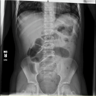
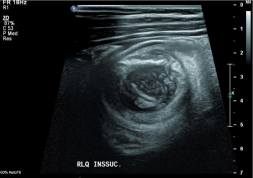
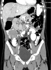
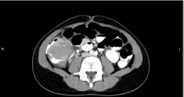
A plain abdominal radiograph was obtained, which showed dilated small bowel loops in the mid- abdomen with findings suggestive of obstruction (Figure 1). An abdominal ultrasound showed telescoping bowel loops and a concentric mass (Figure 2). A CT scan of the abdomen and pelvis showed a homogeneous mass/filling defect within the cecum (Figure 3A and B). The operating findings included copious peritoneal fluid with ileocolic intussusception with dilated terminal ileum but viable bowel. There were also a cecal mass, enlarged mesenteric lymph nodes and a normal appendix. An exploratory laparotomy and ileocecectomy with primary ileocolostomy was performed. The surgical pathology report identified the abdominal mass asa Burkitt Lymphoma. Patient was then referred to a cancer center for further management.

Figure 1: Intussusception CR (Xray).

Figure 2: Intussusception CR (US).

Figure 3a: Intussusception CR (CT).

Figure 3b: Intussusception CR (CT).
Discussion
We report the case of a 9-year-old boy with intussusception secondary to an intra-abdominal Burkitt lymphoma. The intussusception of this patient was a pathology occurring outside of the typical age range of 5 months to 2 years of age [1-5]. The initial presentation of intussusception is non-specific. [1-6] Abdominal pain can be due to a large variety of clinical conditions [1-5]. In older children conditions with pathologic lead points should be considered [1,4]. Pathologic lead points can range from lymph nodes to tumors, this is why it is important to identify them and rule out any malignancies [1-4,7]. A 22% incidence of lead points has been reported in children older than 2 years of age [1]. These pathologic lead points are considered the main causes of intussusceptions [1-4,8,10]. The common presenting symptoms, regardless of the etiology of the intussusception, tend to be vomiting, abdominal pain, distention, diarrhea or other non-specific symptoms [1-4,7]. Symptoms can vary in frequency and can also vary depending on the age at presentation [1-4]. A “sausage shaped mass” has been described on physical exam along with a right lower quadrant that is scaphoid. However this cannot always be palpated [1-4,6,7]. The above mentioned symptoms can be related to a great number of conditions ranging from “acute gastroenteritis” to surgical conditions like appendicitis and intussusceptions [6]. This is why it is important to recognize and identify those that can be life threatening early in order to prevent complications [1,2,6].
As we can see in this patient; diagnosis of intussusception may be difficult when the symptoms are non-specific. This patient required multiple ED visits and a wide variety of diagnostic methods to identify the cause of the abdominal pain. Many different tools may be used to get a proper diagnosis, as symptoms can be non-specific and can be episodic in nature leading to normal physical exams at the time of evaluation [1,8,9]. Abdominal imaging can range from x-rays, point-of-care ultrasound, abdominal CT, and formal ultrasounds [1,8,9]. This case highlights the importance of a proper diagnosis. Since missing it could have not only caused bowel necrosis; but, could have led to a spread of the mass if left untreated.
Ultrasound is the initial diagnostic test of choice, since it can now be performed bedside in the ED [1]. It is important to remember that the treatment of intussusception is different if it is caused by a PLP. Thus surgery will more likely be required to remove the cause [1,9]. While first line treatment for intussusception usually is in the form of an air or hydrostatic enema, this treatment could still lead to recurrence in our patient due to the underlying lymphoma [1,3,4,6,7]. Different diagnostic methods must be employed if necrosis of the tissue is suspected, or as in this case there is a PLP causing the intussusceptions [1,3,6,7]. Causes of intussusception tend to be idiopathic between 80-90%. Nevertheless, malignancies should always be considered for in patients older than 2 years of age. Children with secondary causes are difficult to reduce and are at higher risk of recurrence. This case underscores the need to consider secondary causes of intussusception in older children. So, we are reminded while uncommon, it should still be kept on our list of differential diagnosis when working up a patient. Any delay in diagnosis may adversely affect the outcomes of patients and this case highlights why an early diagnosis is so important. Several factors have been identified that may predict recurrent intussusception. These include age (i.e.,over 1 year of age), symptom duration for more than12 hours, the absence of vomiting, mass location (right abdomen) and pathological lead points [11].
This report underscores the need of intussusception outside of the typical age range, should raise the suspicion of a lead point. Lead points are of concern since there is a high risk of malignancy, and the longer they are left untreated the greater is the risk to the patient. It should be noted that the classic triad of intermittent abdominal pain, blood in the stool and vomiting may not be present on initial presentation. This case highlights the importance of the recognition of pathologic lead points as the management of the patient will vary with the cause of the intussusception. Henceearly recognition and prompt treatment can help to improve the outcome.
References
- Maqbool A, Liacouras CA. Closed-Loop Obstructions. Published online 2021. doi:10.1016/B978-0-323-52950-1.00359-X
- Ye X, Tang R, Chen S, Lin Z, Zhu J. Risk factors for recurrent intussusception in children: A systematic review and meta-analysis. Front Pediatr. 2019; 7: 1-8.
- Beasley SW. The ‘ins’ and ‘outs’ of intussusception: Where best practice reduces the need for surgery. J Paediatr Child Health. 2017; 53: 1118-1122.
- Charles T, Penninga L, Reurings JC, Berry MCJ. Intussusception in children: A clinical review. Acta Chir Belg. 2015; 115: 327-333.
- Stringer MD, Pablot SM, Brereton RJ. Paediatric intussusception. Br J Surg. 1992; 79: 867-876. doi:10.1002/bjs.1800790906
- Waseem M, Rosenberg HK. Intussusception. 2008; 24: 793-800.
- Edwards EA, Pigg N, Courtier J, Zapala MA, MacKenzie JD, Phelps AS. Intussusception: past, present and future. Pediatr Radiol. 2017; 47: 1101- 1108.
- Binkovitz LA, Kolbe AB, Orth RC, et al. Pediatric ileocolic intussusception: new observations and unexpected implications. Pediatr Radiol. 2019; 49: 76- 81.
- Lin X kun, Xia Q zhang, Huang X zhong, Han Y jiang, He G rong, Zheng N. Clinical characteristics of intussusception secondary to pathologic lead points in children: a single-center experience with 65 cases. Pediatr Surg Int. 2017; 33: 793-797.
- Wassmer CH, Abbassi Z, Ris F, Berney T. Intussusception in an immunocompromised patient: A case report and review of the literature. Am J Case Rep. 2020; 21: 1-7.
- Guo WL, Hu ZC, Tan YL, Sheng M, Wang J. Risk factors for recurrent intussusception in children: A retrospective cohort study. BMJ Open. 2017; 7: 1-6.