
Case Report
Austin J Clin Case Rep. 2022; 9(6): 1264.
Application of Kinesio Taping Method on a Stroke Patient with Hemiplegic Shoulder Pain: a Case Report
Kwong Ming-kin* and Alice Po Shan Chan
Occupational Therapy, Tai Po Hospital, Hong Kong
*Corresponding author: Kwong Ming-kin, Occupational Therapy, Tai Po Hospital, Hong Kong
Received: September 29, 2022; Accepted: October 26, 2022; Published: November 02, 2022
Abstract
Kinesio Taping (KT) method is a potential alternative to enhance treatment outcomes for stroke patients with hemiplegic shoulder pain. A 69-year-old man with a hemorrhagic stroke was presented in this case report to explore the effect of KT method in optimizing functional outcome of hemiplegic upper limb in stroke patients. Seven days of KT method as an adjunct to conventional OT training was provided and results showed clinically significant improvement in ADL function and hemiplegic upper limb function. KT method has been shown to be effective in alleviating pain and increasing passive range of motion. Yet, further studies of a larger scale would warrant its treatment effectiveness in the stroke population.
Keywords: Kinesio taping; Tape; stroke; Shoulder pain; Occupational therapy
Introduction
Stroke is one of the most disabling neurological diseases with up to 84% of post-stroke patients experiencing Hemiplegic Shoulder Pain (HSP) [1]. HSP is one of the major causes of reduced upper limb function, which accounts for 70-80% of the post-stroke population [2,3]. Rehabilitation progress could be hindered by the HSP, which limits patients’ performance in Activities of Daily Living (ADL) and reduces the quality of life [3].
There are various proposed causes of HSP, including glenohumeral subluxation, impingement, rotator cuff tear, adhesive capsulitis, spasticity, neuropathic factor, etc. Researchers tend to conclude with a multifactorial etiology [4]. However, Peters found no significant relationship between glenohumeral subluxation and HSP [5]. In contrast, Walsh observed that HSP did not occur until spasticity developed [6]. Evidence also showed that HSP had a significant correlation with spasticity and limited Range of Motion (ROM) [11]. Spasticity, therefore, may play a significant role in HSP when compared to other proposed etiologies.
Traditional non-pharmacological interventions for HSP include active or passive mobilization, shoulder support, neuromotor techniques, electrical stimulation, etc.,[5]. Occupational therapists provide functional training after the patient’s hemiplegic shoulder has been prepared by the above interventions [8]. However, the above interventions are often time-consuming with uncertain efficacy [5]. Moreover, spasticity and HSP are often triggered by poor handling and positioning beyond Occupational Therapy (OT) sessions. Therefore, a less time-consuming intervention that can continue normalizing muscle tone after an OT session is preferred.
Kinesio Taping (KT) method could be utilized in preparation procedures to optimize the efficiency and effectiveness of each OT session. Kenzo Kase, the creator of KT method, claimed an application of Kinesio tape can improve circulation and reduce pain by restoration of superficial and deep fascia function [9]. Application targeting muscle layers can alter muscle tone and, therefore, can potentially reduce HSP. Kinesio tape can be worn for a long period of time to continue treatment beyond treatment sessions [10]. A pilot study found that Kinesio tape showed no effect on HSP and ROM [11] while another randomized study found a potential effect in the acute phase [12]. Nevertheless, these studies applied the same taping design to all subjects without individual assessment.
In this report, a single case is presented to explore the effectiveness of application of KT method in maximizing treatment outcomes of the hemiplegic upper limb in a stroke patient.
Case Report
The Subject
A 69-year-old man, Mr. Z, with a past medical history of hypertension, suffered from intracerebral hemorrhage at left basal ganglia and was admitted to the hospital. Mr. Z presented with right hemiplegia and slurred speech. After two weeks of acute hospital stay, Mr. Z was transferred to the rehabilitation hospital. During the initial assessment, he complained of right shoulder pain with onset after his admission to the rehabilitation hospital. The function of his right upper limb and the passive ROM of his right shoulder was impaired. There was spasticity over shoulder flexors as well as flexor muscles in the right upper limb, without shoulder subluxation. His HSP was suspected to be caused by spasticity.
Assessment
Mr. Z was assessed using the following assessment protocol during the 1-week intervention period (Table 1).
![]()
    Measuring Parameters

    Assessment

    Assessment Frequency


    Hemiplegic shoulder pain

    Pain VAS

    Daily


    Passive ROM

    SROMP

    Daily


    Hemiplegic upper limb function

    FTHUE-HK

    Pre and Post intervention


    MESUPES-arm

    Pre and Post intervention


    ADL performance

    mBl

    Pre and Post intervention
Table 1: The 1-week assessment protocol.
Shoulder pain and passive ROM were measured daily using Pain Visual Analogue Scale (VAS) and Shoulder lateral rotation ROM measured at the point of Pain (SROMP) [13] respectively. For SROMP, the subject lies in a supine position with 45-degree shoulder abduction and 90-degree elbow flexion. Measurement is taken when the subject reports pain during passive shoulder lateral rotation.
In addition, the hemiplegic upper limb function and ADL performance were assessed using the Hong Kong version of the Functional Test for the Hemiplegic Upper Extremity (FTHUE-HK), the arm-subset of the Motor Evaluation Scale for Upper Extremity in Stroke Patients (MESUPES-arm) and the Modified Barthel Index (MBI). FTHUE-HK evaluates the ability of hemiplegic upper limb in performing ADL [14]. The limb function is graded from level 1, nonfunctional, to level 7, the highest level. MESUPES-arm takes quality of movement in ADL into account [15]. MESUPES-arm consists of 8 functional tasks. Each task is rated from 0 to 5 according to the quality of movement during the task. A full score of 40 represents the highest hemiplegic upper limb function. The MBI, with a full score of 100, is widely used to assess ADL performance.
Interventions
KT method was applied as an adjunct to conventional OT stroke rehabilitation program for 1 week. At the beginning of each OT session, Mr. Z was assessed by a Certified Kinesio Taping Practitioner. Then, a tape with a tailor-made cut and tension corresponding to the assessment result was applied. Only one tape was applied in each training session. Mr. Z performed ADL and upper limb functional training with the tape in-situ. To prevent improper handling of the tape and unattended allergic responses to the tape, the tape was removed after each OT session.
Result
A week of KT method used as an adjunct to conventional OT treatment was completed. There was no allergy or other adverse response to the tape noted during the intervention period.
Several taping methods were applied (Table 2). One of the assessment results indicated that Mr. Z’s right shoulder passive ROM was improved by providing space to the anterior deltoid. Thus, one web-shaped tape was applied to the anterior deltoid with space correction technique. According to assessment results on another treatment session, shoulder pain during passive ROM can be reduced by manually gliding the pectoralis major muscle distally. Therefore, a Y-shaped tape was applied to the pectoralis major with overactive muscle correction technique to reduce the muscle activity of the hypertonic muscle (Figure 1).
![]()
    Day

    KT technique

    Target tissue

    Cut


    1

    Overactive muscle correction

    Pectoralis major

    Y-shaped tape


    2

    Overactive muscle correction

    Pectoralis major

    Y-shaped tape


    3

    Fascia correction

    Fascia over anterior deltoid

    Small web-shaped tape


    4

    Space correction

    Anterior deltoid

    Web-shaped tape


    5

    Overactive muscle correction

    Supraspinatus

    I-shaped tape
Table 2: KT techniques over the 1-week intervention period.
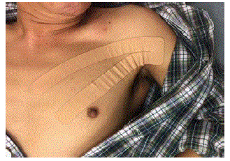
Figure 1: A Y-shaped tape over the pectoralis major with overactive muscle correction technique.
After each treatment session, there was a reduction in shoulder pain VAS and an increase in SROMP [Figure 2]. Over the one-week treatment, the shoulder pain VAS showed a reducing trend with an overall 60% reduction in pain. The SROMP also showed an increasing trend with an overall 46% improvement. In addition, the FTHUEHK, MESUPES-arm and MBI also improved after the one-week intervention period (Table 3).
![]()
    Pre

    Post

    Percentage change


    FTHUE-HK

    3

    4

    -


    MESUPES - Arm

    11

    10

    172%


    mBl

    31

    43

    39%
Table 3: Functional outcomes of the 1-week KT method as an adjunct to

conventional OT training.
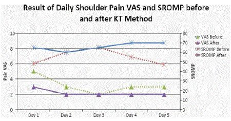
Figure 2: Effect of KT method on shoulder pain and shoulder ROM.
Discussion
This case report aimed to explore the effect of KT method in optimizing OT treatment outcomes. The results showed an immediate pain-relieving effect and improvement in passive ROM in shoulder lateral rotation by KT method. With these effects, the patient’s shoulder joint had already been prepared for further upper limb functional training and ADL training with the tape in-situ. Moreover, the pain-relieving effect and the improvement in the shoulder passive ROM were carried over to the next OT treatment session. Both ADL performance and upper limb function were shown to have clinically improved.
Preparation of affected joints is crucial for upper limb functional training in OT stroke rehabilitation. Compared to traditional timeconsuming preparation procedures, Kinesio tape provides immediate effects on pain relief and improving shoulder ROM. Having the KT method as an adjunct to conventional OT training, more time could be spared for upper limb functional training and ADL training, and therefore optimizing resource utilization and OT treatment outcomes.
It is believed that KT produces an immediate pain-relieving effect by altering hemiplegic muscle tone. A similar pain-relieving effect is also produced in KT assessment by distally gliding the pectoralis major muscle. Moreover, the Y-shaped tape was applied to the chest but not directly to the shoulder. The pain-relieving effect on the shoulder could hardly be explained by improved circulation. Restoration of fascia function could also be another possible mechanism. Further laboratory investigation on the effect of KT on fascia function is suggested.
The carryover effect of Kinesio tape is demonstrated in this case report despite the limited time for tape application. KT method suggests that the tape can be kept on skin for 3 days. In hospital settings, collaboration with multidisciplinary team members would be necessary in order to avoid improper handling and unattended allergic responses. Further exploration on the applicability of KT method in various clinical conditions as well as better communication with other members in the multidisciplinary team is indicated.
There was no control subject in the present study. Besides, there was a lack of statistical evidence to support the effect of KT method on long term treatment outcomes. Future larger scale randomized controlled trial study on the effectiveness of KT method is indicated.
Conclusion
This case report documents the immediate effect of KT method on relieving HSP in a post-stroke patient. Together with conventional OT treatment, a week of Kinesio tape application can lead to improvements in upper limb function and ADL performance. The application of KT method on stroke patients with HSP as an adjunct to conventional OT treatment is promising.
Declaration
The authors declare that there is no conflict of interest.
References
- Pillastrini P, Rocchi G, Deserri D, Foschi P, Mardegan M, et al. Effectiveness of neuromuscular taping on painful hemiplegic shoulder: a randomised clinical trial. Disabil Rehabil. 2015; 38: 1603-1609.
- Shah MV, Kumar S, Muragod AR. Effect of Constraint Induced Movement Therapy v/s Motor Relearning Program for Upper Extremity Function in Sub Acute Hemiparetic Patients-a Randomized Clinical Trial. Indian Journal of Physiotherapy and Occupational Therapy - An International Journal. 2016; 10: 71.
- Sommerfeld DK, Eek EUB, Svensson AK, Lotta Widén H, Arbin MHV. Spasticity After Stroke. Stroke. 2004; 35: 134-139.
- Kashi Y, Ratmansky M, Defrin R. Deficient Pain Modulation in Patients with Chronic Hemiplegic Shoulder Pain. Pain Pract. 2018; 18: 716-728.
- Peters SB, Lee GP. Functional Impact of Shoulder Taping in the Hemiplegic Upper Extremity. Occup Ther Health Care. 2003; 17: 35-46.
- Walsh, K. Management of shoulder pain in patients with stroke. Postgrad Med J. 2001; 77: 645-649.
- Namdari S, Alosh H, Baldwin K, Mehta S, Keenan MA. Shoulder tenotomies to improve passive motion and relieve pain in patients with spastic hemiplegia after upper motor neuron injury. J Shoulder Elbow Surg. 2011; 20: 802-806.
- Jarvis K, Reid G, Edelstyn N, Hunter S. Development of the Occupational Therapy Stroke Arm and Hand Record: An Upper Limb Treatment Schedule. British Journal of Occupational Therapy. 2014; Z7: 126-133.
- Williams S, Whatman C, Hume PA, Sheerin K. Kinesio Taping in Treatment and Prevention of Sports Injuries. Sports Med. 2012; 42: 153-164.
- Kase K, Hashimoto T, Okane, T. Kinesio Taping Perfect Manual: Amazing Taping Therapy to Eliminate Pain and Muscle Disorders. Kinesio USA. 1998.
- Kalichman L, Frenkel-Toledo S, Vered E, Sender I, Galinka T, et al. Effect of kinesio tape application on hemiplegic shoulder pain and motor ability. Int J Rehabil Res. 2016; J9: 272-276.
- Huang Y, Chang K, Liou T, Cheng C, Lin L, et al. Effects of Kinesio taping for stroke patients with hemiplegic shoulder pain: A double-blind, randomized, placebo-controlled study. J Rehabil Med. 2017; 49: 208-215.
- Bohannon RW, Andrews AW. Shoulder Subluxation and Pain in Stroke Patients. Am J Occup Ther. 1990; 44: 507-509.
- Fong K, Ng B, Chan D, Chan E, Ma D, et al. Development of the Hong Kong Version of the Functional Test for the Hemiplegic Upper Extremity (FTHUEHK). Hong Kong Journal of Occupational Therapy. 2004; 14: 21-29.
- Winckel AVD, Ehrlich-Jones L. Measurement Characteristics and Clinical Utility of the Motor Evaluation Scale for Upper Extremity in Stroke Patients. Archives of Physical Medicine and Rehabilitation. 2018; 99: 2657-2658.