
Case Series
Austin J Clin Case Rep. 2022; 9(6): 1266.
Is Polychondritisa Form of IBD Extraintestinal Manifestation? A Case Series of Patients with Concomitant Relapsing Polychondritis and Inflammatory Bowel Disease
Azar E*, Goldin E and Shitrit ABG
Department of Gastroenterology, Shaare Zedek Medical Center, Israel
*Corresponding author: Ariella Bar-Gil Shitrit, Department of Gastroenterology, Shaare Zedek Medical Center, Israel
Received: October 28, 2022; Accepted: November 23, 2022; Published: November 30, 2022
Abstract
Some patients with Inflammatory Bowel Disease (IBD) may experience manifestations in sites other than the gastrointestinal tract. Those Extraintestinal Manifestations (EIMs) may have special treatment projections for those patients.
Relapsing Polychondritis (RPC) is a rare auto inflammatory disease that can involve any cartilage tissue of the body, most commonly the cartilages of the ear, nose and eyes but can also affect major airways and the heart. While some cases take more of a benign course with mild to moderate symptoms, some patients will experience devastating debilitating symptoms and even death. Little is known about the pathogenesis of the disease and there are no guidelines for treatment, although immunomodulatory and anti-inflammatory treatment had been described in the literature.
In this case series we describe the history of 4 patients with a diagnosis of both RPC and IBD, suggesting a connection between the diseases and the hypothesis that polycondritis is a form of EIM. Acknowledging this may lead to more cases being revealed and to better understanding and more effective treatments.
Keywords: Inflammatory bowel disease; Crohn’s disease; Ulcerative colitis; Extraintestinal manifestations; Relapsing polychondritis
Background
Inflammatory Bowel Disease (IBD) consists majorly of two disorders: Crohn’s Disease (CD) and Ulcerative Colitis (UC) and affect millions of people worldwide, mostly in industrialized countries [1]. IBD affects not only the gastrointestinal tract but also other organs of the body, commonly referred to as Extra Intestinal Manifestations (EIMs) of IBD. EIMs has been reported to affect 6-47% of IBD patients. EIM most commonly affect the musculoskeletal system followed by dermatologic, ocular and hepatobiliary involvement. Other, less frequent, forms of EIM can involve the lungs, heart, pancreas or vascular systems [2]. EIMs can present before or after first diagnosis of IBD [3] and can be related, or not, to disease activity. IBD is a chronic and insidious disease that present many challenges for the patient and his treating physician and EIMs are another layer of complexity to the situation and may affect treatment choices and therefore should be recognized accordingly.
Relapsing Polychondritis (RPC) is a rare autoimmune disease characterized by recurrent, episodic inflammation of cartilaginous tissues. The annual incidence of RPC is 0.7-3.5:1,000,000. The disease severity may vary from mild occasional symptoms to a debilitating, sometimes life-threatening disease. Involvement of the auricular cartilage is the most common manifestation. Unilateral or bilateral external ear inflammation that doesn’t involve the ear lobe is the typical auricular manifestation. Other involved cartilage tissues can be those of the nose, eyes, joints, heart (mostly the aortic and mitral valves), large airways and kidneys. About one third of patients present with other diseases, most commonly systemic vasculitis. While in some cases the disease can be of benign fashion causing mild local symptoms, some patients may experience a more aggressive form that can lead to disabilities such as deafness or blindness and in some cases can lead to death, most commonly by airways and heart involvement. Diagnosis of RPC is based on clinical features and few diagnostic criteria have been suggested. Accurate assessment of the overall prognosis of the disease is difficult because of the disease rarity and reporting bias [4,5]. There are no clinical guidelines for RPC and for treatment of RPC. Treatment of mild cases consists of Non-Steroidal Anti-Inflammatory Drugs (NSAIDs) or dapsone. More severe cases are treated with systemic corticosteroids, especially when cardiac, ocular or airway involvement is apparent. Long term steroid treatment does not appear to change to course of the disease and severe cases is frequently treated with immunomodulators such as cyclophosphamide, cyclosporine, azathioprine and methotrexate. With the advancements of biologic treatments in recent years, patients were reported to be treated with biologic treatments such as TNFa-inhibitors, tocilizumab, anakinra, rituximab and abatacept [6].
A connection between RPS and IBD has been described in few case studies of patients with UC [7-10] and CD [11-13]. Here we describe 4 patients exhibiting both IBD and RPC.
Case 1
A 33 year old woman presented with a two year history of abdominal pain, diarrhea and weight loss. Initial evaluation included gastroscopy and colonoscopy which was normal and borderline fecal calprotectin results (177 ug/gr), Computed Tomography (CT) enterography showed signs of terminal ileitis and she started treatment with systemic steroids, Azathioprine and 5-ASA but showed no benefit. Due to diagnostic uncertainty those treatments were stopped. She was then re-evaluated and colonoscopy showed mucosal erythema of terminal ileum with some aphtous ulcers. Abdominal US showed thickening of ileocecal valve, Magnetic Resonance imaging Enterography (MRE) was normal but endoscopic capsule showed diffuse ulcerations and aphtous ulcers and CD was diagnosed.
About a year prior to her first gastrointestinal symptoms she was admitted due to nose inflammation that appeared as bilateral cellulitis with septum inflammation demonstrated by CT. She was treated with augmentin and responded slowly. Two years after, she was admitted again with right auricular perichondritis with no improvement under a course of topical treatment and later a course of cephalexin. She was then diagnosed with RPC and treated with oral prednisone and responded quickly.
After she was diagnosed with CD she was planned to start treatment with infliximab but didn’t because of personal reasons. She was then admitted again for auricular perichondritis, this time of her left ear and was treated with oral prednisone with improvement of both her abdominal and auricular symptoms. After symptoms subsided she started treatment with adalimumab and stayed in remission. She had her last episode of perichondritis 3 years later while she wasn’t taking adalimumab for 3 months for causes of nonadherence and was, again, treated successfully with oral prednisone.
Case 2
A 29 year old woman, with history of psoriasisas well as recurrent vulvovaginitis and condyloma, was first presented to us with 1 year history of diarrhea, abdominal pain and bloating. She was with no active treatment for psoriasis. Colonoscopy was normal and gastroscopy showed chronic gastritis, negative to Helicobacter Pylori. Endoscopic capsule showed gastritis, duodenitis and jejunitis without ileal involvement.
She had a first event of perichondritis of auricle and otitis externa of her right ear about 2 years prior to her appearance at our clinic. She was admitted and treated with oral ciprofloxacin and topical treatment and improved. Her second event was of perichondritis of her left auricle, again she was treated with antibiotics – ciprofloxacin and cefazolin and improved again. After those events she developed Immune Thrombocytopenia Purpura (ITP) and due to mucosal bleeding started treatment with oral prednisone and later with azathioprine and tapering down of prednisone until fully stoppage. She did have another flare of ITP under azathioprine treatment and the treatment was stopped. She needed another course of prednisone. Meanwhile she didn’t experienced other episodes of perichondritis but about 6 months after she stopped the steroidal treatment she was admitted again with bilateral otitis externa and nasal cellulitis. At that point she was diagnosed with RPC and went through a rheumatologic evaluation whichruled out other systemic diseases (eg. Behcet syndrome, systemic lupus erythematosus, hepatitis B and C, human immunodeficiency virus, vasculitis and connective tissue disease). She was treated with antibiotics (ciprofloxacin and cefazolin) and topical treatment and seemed to be improving but later had another episode of perichondritis of her right ear that went along with exacerbation of psoriasis. At that point, after multidisciplinary discussion she started treatment with Infliximab. She received infliximab but after receiving 3 doses she experienced an exacerbation of Crohn’s, without events of chondritis. Due to her history of psoriasis she started treatment with Ustekinumab for both IBD and psoriasis and had a good response but 3 months later was admitted twice for events of bilateral auricle chondritis and nasal tip chondritis, both treated successfully with oral prednisone.
Case 3
A 39 year old woman presented 11 years ago with perichondritis of her right pinna with slow improvement under antibiotics – oralaugmentin at first and later ciprofloxacin and cefazolin. She also reported previous inflammation of nose-bridge that passed with oral antibiotic treatment and a few episodes of episcleritis / scleritis. In the months following she developed mouth aphtous ulcers and erythema nodosum. A suspected diagnosis of Behcet syndrome was made but she never met full criteria, and treatment with oral prednisone and methotrexate was initiated. Six months later she started complaining of bloody diarrhea. Colonoscopy demonstrated pancolitis with diffuse ulcerations. Biopsies were inconclusive to determine between Behcet syndrome and IBD and showed ulcerations with mononuclear infiltration without granulomas. Since she didn’t comply with diagnostic criteria for Behcet, a diagnosis of CD was made. As she was still symptomatic she started Azatioprine as steroid-sparing treatment but relapsed so she started Adalimumab as well and was able to wean from systemic steroid treatment and was stable with no complaints under treatment with Adalimumab and Azatioprine as described. Four years later she was still asymptomatic, she stopped treatment of adalimumab because of pregnancy and since then she is asymptomatic under Azatioprine as sole treatment for 6 years.
Case 4
A 63 year old woman, with 12 years history of CD. she was first diagnosed after screening colonoscopy showed focal regions of erythema, edema and ulcerations over the right and transverse colon. Biopsy showed chronic active crypt-destructive colitis. The patient had history of arthritis and anterior uveitis. Since she had no gastrointestinal symptoms she was first treated with 5-ASA. Four years after the diagnosis she became symptomatic with bloody diarrhea and weight loss but reclined corticosteroid treatment. Six months after, she was diagnosed with auricular perichondritis that was not responsive to a 10 days antibiotic course but responded to oral prednisone course with tapering-down regimen. Later she had exacerbation of her CD with endoscopic involvement of her rectum and descending colon and simultaneously she had an event of uveitis which required oral corticosteroid treatment. She was then started on anti-TNF treatment with adalimumab and since then is in remission with no recurrences.
Discussion
A connection of IBD and RPC to date was demonstrated with few case reports [7-13]. In this case series we report 4 cases of patients who presented with both IBD and RPC. The clinical data of the patients is summarized in (Table 1). All patients were diagnosed with RPC after meeting the modified McAdam’s criteria [14]. The rarity of RPC give rise to the question about a possible connection between the two diseases or that the two share common characteristics. Both diseases are considered to be auto inflammatory and may share common inflammatory pathways and share common treatments.
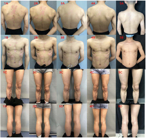
    Figure 1: Week 0 of treatment; Figure 2: Week 1 of treatment; Figure 3: Week 2 of treatment; Figure 4: Week 4 of treatment; Figure 5: Week 8 of treatment.

Note: A: Back view of trunk; B: Frontal view of trunk; C: Frontal view of lower extremily; D: Back view of lower extremity.
![]()
    Case #

    IBD type

    Sex

    Age

    RPC organs involvement

    Treatments


    1

    CD

    F

    33

    Nose, both ears

    PRD, ADA


    2

    CD

    F

    29

    Both ears, nose

    PRD, AZA, IFX, UST


    3

    CD

    F

    39

    Nose, right ear, eyes

    PRD, AZA, ADA


    4

    CD

    F

    63

    Eyes, right ear, joints

    PRD, ADA


    CD: Crohn's Disease; F: Female; PRD: Prednisone;    AZA: Azathioprine; ADA: Adalimumab; IFX: Infliximab; UST: Ustekinumab
Table 1: Clinical characteristics of patients with inflammatory bowel disease and

relapsing polychondritis.
In this study we found a cluster of patients exhibiting symptoms and the diagnosis of both diseases. While awareness for IBD is high in the medical community and in the general population, RPC is less known and may go under diagnosed and underreported by patients and physicians, and therefore mistreated. More study should be done to further demonstrate this relation between RPC as an EIM of IBD.
References
- Ng Siew C, Shi HY, Hamidi N, Underwood FE, Tang W, et al. Worldwide incidence and prevalence of inflammatory bowel disease in the 21st century: a systematic review of population-based studies. The Lancet. 2017; 390: 2769-2778.
- Vavricka Stephan R, Schoepfer A, Scharl M, Lakatos P, Navarini A, et al. Extraintestinal manifestations of inflammatory bowel disease. Inflammatory bowel diseases. 2015; 21: 1982-1992.
- Vavricka Stephan R, Rogler G, Cantenbein C, Spoerri M, Vavricka MP, et al. Chronological order of appearance of extraintestinal manifestations relative to the time of IBD diagnosis in the Swiss inflammatory bowel disease cohort. Inflammatory bowel diseases. 2015; 21: 1794-1800.
- Trentham David E, Christine H Le. Relapsing polychondritis. Annals of internal medicine. 1998; 129: 114-122.
- Borgia Francesco, Giufrrida R, Guarneri F, Cannavo SP. Relapsing polychondritis: an updated review. Biomedicines. 2018; 6: 84.
- Grygiel-Górniak Bogna, et al. Relapsing polychondritis: state-of-the-art review with three case presentations. Postgraduate medicine. 2021; 133: 953-963.
- Rosen SW, Mackenzie MR, Cohen PJ, Friedlander S, Medart W, et al. A syndrome resembling relapsing polychondritis in a patient with ulcerative colitis. Gastroenterology. 1969; 56: 323-330.
- Ueno Y, D Chia, EV Barnett. Relapsing polychondritis associated with ulcerative colitis. Serial determinations of antibodies to cartilage and circulating immune complexes by three assays. The Journal of rheumatology. 1981; 8: 456-461.
- Almadi Majid A, AlEnizi AT, Menard HA, Hizenrat N. Relapsing polychondritis and ulcerative colitis. Saudi journal of gastroenterology: official journal of the Saudi Gastroenterology Association. 2010; 16: 49.
- Hanioka Y, Shimizu K, Yamagami K, Yao S, Nakamura R, et al. Exacerbation of Ulcerative Colitis with Tocilizumab: A Report of Two Cases, One with Takayasu Arteritis and the Other with Relapsing Polychondritis. Internal Medicine. 2021; 60: 1615-1620.
- Touma DJ, Gross EJ, Karmody CS, Fawaz KA. Relapsing polychondritis in association with Crohn’s disease. American journal of otolaryngology. 1996; 17: 424-426.
- Jung Hye-In, Kim HJ, Kim JM, Lee JY, Park KS, et al. Co-existence of relapsing polychondritis and Crohn disease treated successfully with infliximab. Yeungnam University Journal of Medicine. 2021; 38: 70-73.
- Naidu H, Szeto W, Kissin E, Farraye FA. MAGIC Syndrome in a Patient with Crohn’s Disease. Inflammatory bowel diseases. 2018; 24: 664-665.
- Damiani Joseph M, Howard L. Levine. Relapsing polychondritis—report of ten cases. The Laryngoscope. 1979; 89: 929-946.