Special Article - Ultrasound Imaging
Austin J Clin Med. 2015;2(1): 1019.
Use of Bedside Ultrasound to Diagnose a Bleeding Hepatic Mass in a Patient with Post-Prandial Right Upper Quadrant Pain
Daniel A Dworkis MD PhD, Kristen Vella Gray PA-C and Sarah E Frasure MD*
Department of Emergency Medicine, Harvard Medical School, USA
*Corresponding author: Sarah E Frasure MD, Department of Emergency Medicine, Brigham and Women’s Hospital Neville House - 236-A, 75 Francis St, Boston, MA 02115, USA
Received: December 05, 2014; Accepted: January 06, 2015; Published: January 19, 2015
Abstract
Emergency physicians can utilize bedside ultrasound to aid them in the diagnosis of a variety of abdominal complaints. In this case an emergency physician utilized bedside abdominal ultrasound to diagnose intra-abdominal free fluid in an elderly patient with post-prandial abdominal pain. Without bedside ultrasound, this patient would have likely been discharged from the emergency department without correctly making a crucial, life saving diagnosis. The sonographic appearance and diagnostic criteria of an abnormal abdominal ultrasound, characterized by intra-abdominal free fluid, are reviewed. Bedside ultrasound by a physician trained in this imaging modality can diagnose abdominal emergencies, facilitating appropriate consultation and treatment.
Keywords: Bedside ultrasound; Right upper Quadrant pain; Hepatic mass
Case Report
A 71 year-old female presented to the Emergency Department (ED) with right upper quadrant (RUQ) pain that developed acutely after the consumption of a cheese burger and fries. She stated that the pain was worse with movement and associated with nausea. By the time she had arrived in the ED her abdominal pain had improved. She denied vomiting, diarrhea, fever, chills, chest or back pain. She had experienced similar RUQ pain approximately one year prior which had subsided without medical intervention. An outpatient abdominal ultrasound ordered by her primary care provider at the time had been unremarkable. Her medical history was notable for hypertension, mitral valve prolapse, and gastroesophageal reflux disease. She took Omeprazole and Metoprolol daily. She denied any history of abdominal surgeries, recent medication changes, and alcohol or illicit drug use.
Vital signs included a blood pressure 150/90 mmHg, heart rate 80 beats per minute, respiratory rate 16 breaths per minute, and temperature of 96.1°F. She had a regular rate and rhythm and clear breath sounds bilaterally. Her abdominal exam was notable for mild tenderness to palpation in the RUQ without associated rebound or guarding. There was no costvertebral angle tenderness to palpation.
The differential diagnosis initially included biliary colic, early cholecystitis, choledocolithiasis, pancreatitis, and gastritis. An electrocardiogram did not demonstrate evidence of ischemia or arrhythmia. Her blood counts were notable for a mild leukocytosis to 14.75 k/μL, a hematocrit of 38%, and platelets of 291 k/μL. Her basic metabolic panel was unremarkable. Her liver function testing results included alanine aminotransferase (ALT) 17 U/L, aspartate aminotransferase (AST) 36 U/L, alkaline phosphatase 65 U/L, and total bilirubin 0.3 mg/dL, all within normal limits. Her lipase level was 36 U/L, also within normal limits. Her urinalysis did not demonstrate evidence of infection.
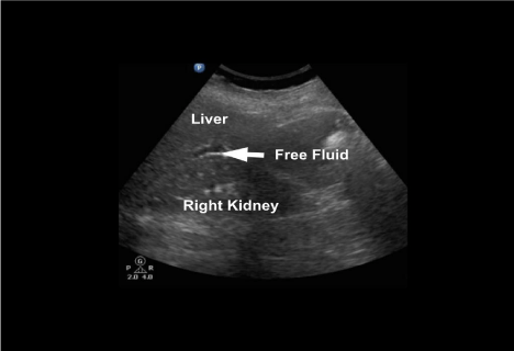
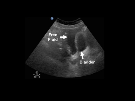
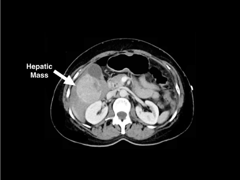
She was provided with Maalox with improvement in her pain and wished to be discharged to attend her granddaughter’s birthday party. The emergency physician (EP) performed a bedside abdominal ultrasound, however, in order to assess for gallstones or evidence of early cholecystitis. Surprisingly, intra-abdominal free fluid was immediately noted in the right upper quadrant (RUQ) and in the suprapubic views (Figures 1 and 2). As a result, an abdominal CT scan was performed which demonstrated a 6.6cm x 5.8cm x 5.4cm mass in the inferior right hepatic lobe with surrounding areas of hemoperitoneum and active hemorrhage (Figure 3). Shortly after the CT scan was obtained, the patient became hypotensive to 80/40 and was resuscitated with a normal saline bolus and one unit of packed red blood cells. Both surgical and interventional radiology consultations were emergently obtained and the patient was taken to the interventional radiology suite, where she underwent embolization of segments 5 and 6 of her right hepatic artery due to active arterial bleeding. Thereafter, she was admitted to the Surgical Intensive Care Unit (SICU) for close monitoring. The following week she underwent a partial hepatectomy with pathology significant for welldifferentiated hepatocellular carcinoma.

    Figure 1:  Right upper quadrant (RUQ) view. Free fluid is noted in Morison’s
Pouch between the liver and kidney (arrow).

Figure 2: Suprapubic view. Free fluid is noted posterior to the bladder.

    Figure 3:  A cross-sectional view of the abdominal CT scan with a large
hepatic mass (arrow).
Hepatocellular Carcinoma (HCC) is a common malignancy whose five year survival rate is less than 12% in the United States [1]. Cirrhosis, which is usually either secondary to a viral infection or prolonged alcohol abuse, remains the strongest risk factor for HCC; greater than 90% of new cases of HCC occur in patients with cirrhotic livers [2]. Current guidelines recommend ultrasound-based surveillance every 6 months to assess for HCC in patients with known cirrhosis [3]. In this case, the patient did not drink alcohol, nor did she have any known risk factors for hepatitis. Furthermore, she had had an abdominal ultrasound one year prior for similar post-prandial pain that had been unrevealing. Given her normal blood work, stable vital signs, and resolving abdominal pain in the ED, she may well have been discharged with a presumptive diagnosis of biliary colic without any imaging studies.
Certainly bedside emergency ultrasound is not specifically designed to evaluate the liver parenchyma. Instead the EP can utilize bedside ultrasound to assess for a variety of diagnoses such as biliary colic, cholecystitis, hydronephrosis, intra-uterine pregnancy, or intraabdominal free fluid [4]. In this case, the EP recognized a moderate amount of free fluid in the RUQ and in the pelvis, and obtained a CT scan, which showed a bleeding hepatic mass. While the true incidence of unexpected liver lesions detected by ultrasound is unknown, population-based screenings in Asian populations suggest a level around 5%, the majority of which are cysts and hemangiomas [5-7]. In this case, a bedside abdominal ultrasound noted intra-abdominal free fluid at the site of her abdominal pain. Further imaging in the ED subsequently identified active extravasation from a hepatic mass that resulted in emergent surgical and interventional radiology consultation, hepatic artery embolization, and SICU admission.
References
- El-Serag HB. Hepatocellular carcinoma. N Engl J Med. 2011; 365: 1118-1127.
- Giannini EG, Cucchetti A, Erroi V, Garuti F, Odaldi F, Trevisani F. Surveillance for early diagnosis of hepatocellular carcinoma: how best to do it? World J Gastroenterol. 2013; 19: 8808-8821.
- Singal A, Volk ML, Waljee A, Salgia R, Higgins P, Rogers MA, et al. Meta-analysis: surveillance with ultrasound for early-stage hepatocellular carcinoma in patients with cirrhosis. Aliment Pharmacol Ther. 2009; 30: 37-47.
- Noble VE, Nelson BP. Manual of Emergency and Critical Care Ultrasound. 2nd Edition. New York, NY, Cambridge University Press. 2011.
- Dietrich CF, Sharma M, Gibson RN, Schreiber-Dietrich D, Jenssen C. Fortuitously discovered liver lesions. World J Gastroenterol. 2013; 19: 3173-3188.
- Rungsinaporn K, Phaisakamas T. Frequency of abnormalities detected by upper abdominal ultrasound. J Med Assoc Thai. 2008; 91: 1072-1075.
- Lu SN, Wang LY, Chang WY, Chen CJ, Su WP, Chen SC, et al. Abdominal sonographic screening in a single community. Gaoxiong Yi Xue Ke Xue Za Zhi. 1990; 6: 643-646.