Clinical Image
Austin J Clin Med. 2018; 5(1): 1035.
Cardiac Lymphangioma in an Adult
Xiaoqian Lu¹, Nan Wang¹, Dianbo Cao² and Yutian Sun¹*
¹Department of Radiology, The First Hospital of JiLin University, Chang Chun
²Pharmaceutical Department, China-Japan Union Hospital of Jilin University, Changchun
*Corresponding author: Yutian Sun, Pharmaceutical Department, China-Japan Union Hospital of Jilin University, Changchun 130033, China
Received: June 11, 2018; Accepted: July 02, 2018; Published: July 09, 2018
Clinical Image
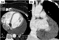
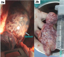
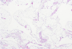
A 39-year-old woman without past medical history was admitted to our hospital for chest discomfort and fatigue on exertional exercise. A large tumoural mass located in the posterolateral wall of the left ventricle was visualized by trans-thoracic echocardiogram. Subsequent chest contrast-enhanced CT showed an unenhanced cystic mass covering the surface of left ventricle, which simultaneously encompassed the left anterior descending artery and concomitant vein (Figure 1). The patient underwent surgery via left thoracotomy under general anesthesia. After opening the parietal pericardium, multicystic dark-purple mass measuring 10.0cm×8.0cm×6.0cm was seen and originated in the left-ventricular cardiac wall with a broad base (Figure 2a). In the process of performing tumor dissection and myocardial suture, cardiac arrest occurred three times and was successfully rescued by cardiac massage and electrical defibrillation. The resected specimen of the tumor revealed a uniformly thin-walled, multicystic lesion with a smooth external surface (Figure 2b) and light yellow serous content. Histological analysis showed a cystic tumor with a predominantly lymphatic vascular component, lymphoid follicles and mature adipose tissue (Figure 3). Final diagnosis was consistent with cardiac lymphangioma. The postsurgical recovery of the patient was uneventful. The lymphangioma is a benign abnormal collection of lymphatic vessels that forms a mass. Most of them occur during childhood and are commonly confined to the head and neck but sometimes may extend into the mediastinum. However, cardiac lymphangioma is exceptionally uncommon and constitutes one of the rare forms of cardiac disease. Because the tumor was found in the left ventricle of the heart and originated in the myocardium in our patient, we performed extensive resection of the tumor together with minimizing the damage to the myocardium.