
Clinical Image
Austin J Clin Med. 2018; 5(2): 1038..
Massive Hemoptysis from Rupture of Aortic Pseudoaneurysm
Lu X¹, Liu Y¹, Cao D¹ and Sun Y²*
¹Department of Radiology, The First Hospital of Jilin University, Chang Chun, China
²Pharmaceutical Department, China-Japan Union Hospital of Jilin University, Changchun, China
*Corresponding author: Yutian Sun, Pharmaceutical Department, China-Japan Union Hospital of Jilin University, Changchun 130033, China
Received: July 03, 2018; Accepted: July 12, 2018; Published: July 19, 2018
Keywords
Aortic Pseudoaneurysm; Complication; Pulmonary Hematoma; CT Angiography
Clinical Image
A 45-year-old male presented with intermittent hemoptysis and progressive dyspnea for 2 days. Chest CT showed a thoracic aortic aneurysm, ill-defined heterogeneous mass in the left upper lobe and patchy attenuation. Moderate pleural effusions were also seen in the left pleural space (Figure 1a-d). The patient refused to surgery and endovascular stent, and died from massive hemoptysis on the 7th day after admission.
Thoracic aortic aneurysm can rupture into adjacent structures, such as pericardial and pleural cavities, trachea and bronchi, esophagus, but rupture into the lung parenchyma is rare. Clinic presentations largely depend on the site of rupture, frequently presenting as sudden death; however, an immediate rupture into the lung, accompanied by fatal hemoptysis and aspiration, is very rarely encountered. CT should be considered the preferred diagnostic technique for the ruptured aneurysm into the lung. CT scan, as described in our case easily detects disseminated alveolar hemorrhage and pulmonary hematoma. Evaluation of the aorta with CT angiography allows confirmation of the aneurysm as well as defining the extent of the diameter.
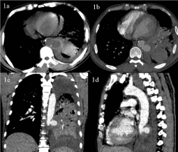
Figure 1: