Clinical Image
Austin J Clin Neurol 2015;2(2): 1022.
Anatomical Variation in DaTscan Imaging Leading to Diagnosis of Cerebral Meningioma
Raul Barahona-Hernando1*, Carlos Ordas- Bandera2, Maria Jose Catalan3 and Eva López- Valdés3
1Department of Neurology, Grupo Hospitalario Quirón, Spain
2Department of Neurology, Hospital Rey Juan Carlos, Spain
3Department of Neurology, Hospital Clínico San Carlos, Spain
*Corresponding author: Raul Barahona, Department of Neurology, Hospital Quiron San Camilo, Juan Bravo 39 St, 28006, Madrid, Spain
Received: January 22, 2015; Accepted: February 10, 2015; Published: February 17, 2015
Editorial
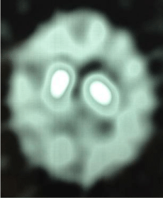
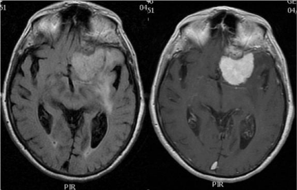
A 79-year-old woman presented with a 3 years history of isolated right-hand tremor. The neurological examination showed intention tremor and mild rigidity in her right hand. Fundoscopic exam showed no papilloedema. The rest of the exam was unremarkable. Single photon emission computed tomography (SPECT) of her brain using the tracer I123-FP-CIT (DaTSCAN) was obtained. Axial sections at the level of the basal ganglia showed homogeneous tracer uptake in both putamen and caudate nuclei. Marked asymmetry in the position of both striatum was noted, with the left one being posteriorly displaced. An MRI was subsequently obtained, that demonstrated a 4,5-cm extraparenchymal space occupying lesion, located at the level of the anterior clinoid process, with marked surrounding edema and homogeneous gadolinium enhacement. The mass compressed the left frontal lobe and displaced the basal ganglia. These findings suggested a meningioma originating from the anterior clinoid process as the most likely differential diagnosis. Conservative management with neuroimaging monitoring control was chosen due to the patient characteristics.
Meningioma may rarely present with Parkinsonism [1-4], and both tracer uptake and absent tracer binding have been reported in DaTSCAN studies obtained in patients with meningioma [3-4]. In our patient, I123-FP-CIT uptake was not demonstrated, but the anatomical variation observed in the SPECT study lead to subsequent neuroimaging that showed the mass, highlighting the importance of careful review of the striatum morphology. Structural neuroimaging is warranted when morphologic abnormalities are found to rule out space occupying lesions.

    Figure 1:  Single photon emission computed tomography using I123-FPCIT
showing homogeneous tracer uptake in both putamen and caudate
nuclei and asymmetry in the position of both striatum, with the left one being
posteriorly displaced.

    Figure 1:  T2 weighted FLAIR MRI showing a lesion with surrounding edema,
and homogeneous gadolinium enhacement on T1-weighted imaging.
References
- Salvati M, Frati A, Ferrari P, Verrelli C, Artizzu S, Letizia C. Parkinsonian syndrome in a patient with a pterional meningioma: case report and review of the literature. Clin Neurol Neurosurg. 2000; 102: 243-245.
- Adhiyaman V, Meara J. Meningioma presenting as bilateral Parkinsonism. Age Ageing. 2003; 32: 456-458.
- Cilia R, Allegra R, Pezzoli G. Meningioma with intense I(123) FP-CIT uptake. Mov Disord. 2012; 27: 1744-1745.
- Biancheri-Mounicq I, Colombié M, Pin JC, Lecoanet A, Adam-Tariel F. Unexpected finding on I-123 FP-CIT SPECT leading to the diagnosis of cerebral meningioma. Clin Nucl Med. 2011; 36: 156-157.