Case Report
Austin J Clin Neurol 2015;2(2): 1024.
Deep Neck Inflammatory Diseases: Implication of Cervical Magnetic Resonance Imaging for Early Diagnosis
Hikichi C, Asakura K, Hirota S, Fukui T, Murate K, Ishikawa T, Kizawa M, Ueda A, Ito S and Mutoh T*
Department of Neurology, Fujita Health University School of Medicine, Japan
*Corresponding author: Mutoh T, Department of Neurology, Fujita Health University School of Medicine, 1-98 Kutsukake-cho, Toyoake, Aichi 470-1192, Japan
Received: January 30, 2015; Accepted: February 18, 2015; Published: February 20, 2015
Abstract
Deep neck inflammatory disorders such as retropharyngeal abscess and pyogenic cervical spondylitis are potentially life-threatening disorders and are quite rare in healthy individuals. With abnormal findings on emergency cervical magnetic resonance imaging (MRI), we succeeded in making prompt diagnoses and initiate appropriate treatments. Both cases recovered almost fully without any orthopedic intervention. Especially in the case of pyogenic cervical spondylitis, we could detect the very early stage of the disease by cervical MRI, i.e., the inflammations were confined to the vertebra without affecting adjacent tissues. Thus, emergency MRI of cervical spine would offer reliable methods for diagnosis and speedy treatments of deep neck inflammatory diseases.
Keywords: Retropharyngeal abscess; Pyogenic cervical spondylitis; Deep neck inflammatory disorder
Introduction
Deep neck infectious disorders in healthy adults (without vertebral surgical history) are very rare but may lead to potentially life-threatening complications [1]. Here, we report two cases, i.e., a case of fulminant retropharyngeal abscess and a case of acute pyogenic cervical spondylitis, both of them showed favorable outcomes. The present cases well illustrated the usefulness and diagnostic importance of emergency MRI examination of the spine.
Case Presentation
Case 1
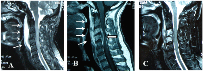
A 39-year-old man had fever and occipital pain. On the next day, he visited a local practice and was prescribed a NSAID and antibiotics. High fever continued following day and occipital pain got worse. He could not turn his neck in any direction. Then, he visited another hospital and was transferred to our hospital. On admission, he was alert. He had a difficulty to open his mouth and had hoarseness. Although other cranial nerves were not involved, nuchal rigidity was observed. Other neurological findings were all normal. Blood examination showed that white blood cell count was 11500 (4000 - 9000/μm3) and CRP was 30.3mg/dl (< 0.3 mg/dl). Cerebrospinal fluid examination disclosed pleocytosis (106 leucocytes/mm3, 82% of lymphocytes) with extremely elevated protein (392 mg/ dl; normal range, 10-40mg/dl) and IgG concentrations (69 mg/ dl; normal range, < 4 mg/dl). Blood culture was negative. PCR examination for tuberculosis in cerebrospinal fluid was negative. The cervical MRI revealed widening of the prevertebral space along C1 to C5 (Figure 1). Under the diagnosis of retropharyngeal abscess, puncture of the abscess and drainage was performed immediately by otolaryngologists. Culture of the abscess was negative, which may be due to the prior administration of antibiotics at other hospital. Previous study has shown that retropharyngeal abscess tends to occur mostly in children and microbiological review of children indicated that anaerobic organisms are predominantly isolated [2]. In adults, aerobic organisms were also isolated [2]. Therefore, a broad spectrum antibiotic (MEMP) was administered intravenously. Eventually, neck pain was alleviated. On 7 days after the diagnosis, however, the patient complained sensory disturbances in his left hand and forearm. Cervical MRI showed the extradural (epidural) abscess in C3 and C4. Therefore, the antibiotic was changed to another broad spectrum antibiotics (PAMP/BP). At 15days after hospitalization, MRI showed prominent shrinkage of extradural abscess. He was discharged at 24 days after hospitalization without orthopedic surgery.

Figure 1: Sagittal cervical MRI of case 1. (A) T2-weighted image on admission. Small arrows indicate the abscess in retropharyngeal space extended from C1 to C5 level. (B) Gadrinium-enhanced T1-weighted image 7 days after admission. Epidural abscess was observed in C3 to C5 level (large arrow). (C) T2-weighted image 15days after admission. Epidural abscess was disappeared and retropharyngeal space was decreased.
Case 2
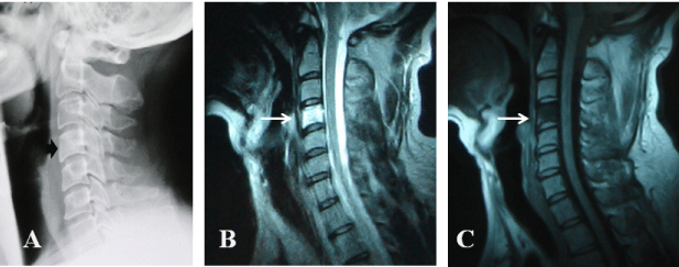
A 47-year-old man gradually developed neck pain in the evening without preceded respiratory tract infection. Moreover, he had no history of vertebral surgery or injuries. Several hours later, he felt severe pain in the neck and left shoulder followed by the difficulties for extending his neck. Anterior chest pain was developed when swallowing. Next morning, he had low grade fever (37.2oC) but had no neck stiffness. Then, he visited our hospital. No muscle weakness or sensory impairment was noted. Laboratory examination revealed mild leukocytosis. CRP was abnormally high, 8.1mg/dl. Erythrocyte sedimentation rate was 72mm/1hour. Although cervical spine X-ray showed no radiographical abnormalities except comparatively hyperintensity as shown in Figure 2A, emergency cervical MRI was performed immediately and it revealed prominent low intensity signal on T1-weighted image and high intensity signal on T2-weighted image in entire C4 (Figure 2B and 2C). The patient was hospitalized and was treated with intravenous administration of antibiotics (CAZ) under the diagnosis of acute pyogenic cervical spondylitis. Neck pain disappeared within a few days. Endoscopic examination of larynx and esophagus did not reveal any abnormalities including fish bones. Blood culture was negative. QuantiFERON-TB test for tuberculosis was negative. Symptoms disappeared and laboratory data were normalized within 10 days after diagnosis. He was discharged at 10 days after hospitalization. He has been followed in our department for more than 3 years and no relapse occurred.

Figure 2: Cervical spine X-ray and sagittal cervical MRI of case 2. Cervical spine X-ray (A), T2-weighted (B), and T1-weighted (C) images on admission. Black arrow indicates mild hyperintensity in C4 (A). White arrows indicate prominent T2 high and T1 low intensity signals in C4 without abscess formation (B and C).
Discussion
Despite the advancement of diagnostic procedures and antibiotics, deep neck infections occasionally cause life-threatening complications due to airway displacement, mediastinitis, spinal cord abscess, jugular vein thrombosis, and carotid artery occlusion [1]. Early diagnosis and treatment are indispensable to prevent complications for not only otolaryngologists but also neurologists.
Retropharyngeal lymph nodes usually disappear after age 4 or 5 years. Therefore, the incidence of non-traumatic retropharyngeal abscess in healthy adults is extremely rare and most of the adult cases are associated with immunocompromised condition or a foreign body complication such as fish bone [3,4]. Due to the rarity of retropharyngeal abscess in adults, the definitive diagnosis may be delayed in some adult patients [5,6]. In our case, foreign body was not identified. Though our first case showed fulminant clinical course with meningeal involvement, swift interdisciplinary treatment with otolaryngologists, i.e., transoral drainage and antibiotics administration were successful.
Pyogenic spondylitis is one of the most severe infectious diseases of the neurological system, and most of the previous cases are related to spinal surgical procedures or tuberculosis infection. Spontaneous pyogenic spondylitis in healthy individuals without any underlying disease is also extremely rare. The cervical spine is a relatively uncommon site for infection in comparison with lumbar and thoracic spine, representing less than 10% of all cases [1]. Pain of shoulder and fever occurred in most of the cases as well as our case 2. Some of them, however, were misdiagnosed initially [7]. Common MRI findings in infectious spondylitis are hypointensity of the involved tissue on T1- weighted images, hyperintensity on T2-weighted images, destruction of two or more adjacent vertebral bodies with involvement of the intervening disc, and epidural and paraspinal extension and/or abscesses [8,9]. We were able to detect the very early stage of the disease confined to C4 vertebral body without affecting adjacent tissues. To our knowledge, no case of pyogenic cervical spondylotis as mild as our case 2 has been reported in the literature.
It should be mentioned that although deep neck inflammatory disorders are quite rare in healthy individuals, detailed and emergent neuroradiological examinations such as MRI are necessary for early diagnosis and treatment of the potentially life-threatening disorders. Our cases highlight the importance of early diagnosis with cervical MRI. With timely diagnosis by cervical MRI, we can initiate the appropriate and intensive treatment of the disorders as seen in the present cases. Fortunately, both present cases recovered almost fully without any orthopedic intervention. These early diagnoses and treatments can help to improve patients' outcome.
References
- Schuler PJ, Cohnen M, Greve J, Plettenberg C, Chereath J, Bas M, et al. Surgical management of retropharyngeal abscesses. Acta Otolaryngol. 2009; 129: 1274-1279.
- Brook I. Microbiology and management of peritonsillar, retropharyngeal, and parapharyngeal abscesses. J Oral Maxillofac Surg. 2004; 62: 1545-1550.
- Dawes LC, Bova R, Carter P. Retropharyngeal abscess in children. ANZ J Surg. 2002; 72: 417-420.
- Page C, Biet A, Zaatar R, Strunski V. Parapharyngeal abscess: diagnosis and treatment. Eur Arch Otorhinolaryngol. 2008; 265: 681-686.
- Schott CK, Counselman FL, Ashe AR. A pain in the neck: non-traumatic adult retropharyngeal abscess. J Emerg Med. 2013; 44: 329-331.
- Popa C, Dekker HM, van Deuren M. Feverless red neck: why worry? Retropharyngeal abscess (RPA) with GABHS. Neth J Med. 2011; 69: 451, 474.
- Feng ZY, Guo F, Chen Z. Literature review and clinical presentation of cervical spondylitis due to salmonella enteritidis in immunocompetent. Asian Spine J. 2014; 8: 206-210.
- Cheung WY, Luk KD. Pyogenic spondylitis. Int Orthop. 2012; 36: 397-404.
- Shousha M, Boehm H. Surgical treatment of cervical spondylodiscitis: a review of 30 consecutive patients. Spine (Phila Pa 1976). 2012; 37: E30-36.