Case Report
Austin J Clin Neurol 2015;2(2): 1026.
Suspected CMV Associated Myelitis in Immunocompetent Adult without Supporting PCR from the CSF
Meir Kestenbaum>1,2, Eitan Auriel E1,2, Haim Perloock2 and Arnon Karni1,2*
1Department of Neurology, Tel Aviv Sourasky Medical Center, Israel
2Sackler’s Medical School, Tel Aviv University, Israel
*Corresponding author: Arnon Karni, Department of Neurology, Tel Aviv Sourasky Medical Center, Tel Aviv University, Tel Aviv, Israel
Received: December 18, 2014; Accepted: March 18, 2015; Published: April 08,2015
Abstract
The occurrence of Cytomegalovirus (CMV) myelitis among immunocompetent patients is extremely uncommon. We present a 53 year old immunocompetent female with myelitis that was associated with infection of CMV that was detected by positive Immunoglobulin M (IgM) antibodies, seroconversion of Immunoglobulin G (IgG) antibodies with low avidity index, but the polymerase chain reaction (PCR) test for CMV in the cerebrospinal fluid (CSF) was negative. We suggest here, that the absence of supporting PCR from CSF or blood CMV antigen should not exclude the diagnosis, especially in the immunocompetent patients.
Keywords: Cytomegalovirus; Cerebrospinal fluid; Computerized tomography; Polymerase chain reaction
Case Presentation
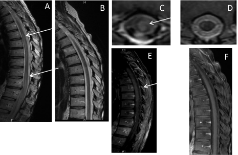
A 53 year old female with no previous significant medical history was hospitalized due to progressive paraparesis. One week prior to her admission she had mild lower back pain with no radiating pain and no neurological impairment. After 5 days she developed a progressive paraparesis combined with lower limbs numbness and urinary incontinence. The patients reported that 2 weeks prior to the onset of symptoms 3 members of the patient’s close family had upper respiratory infection (URI) with positive serology for CMV. The patient had denied any URI symptoms. Neurological exam on admission revealed spastic paraparesis with bilateral extensor plantar response and thoracic (T) sensory level at T8. One day following admission the patient deteriorated and became paraplegic. Laboratory blood tests demonstrated normal complete blood count, C-reactive protein and chemistry with the exception of mild elevation of liver enzymes (alanine aminotransferase, aspartate aminotransferase and Gamma-glutamyl transferase). Computerized tomography (CT) with contrast revealed no significant findings. Magnetic resonance imaging (MRI)of the entire neuro-axis including the T2 weighted images and fluid-attenuated inversion recovery (FLAIR) sequences demonstrated two elongated intramedullary hyperintense lesions at the levels of T5-8 and T9-12 with mild swelling of the cord and enhancement following injection of gadolinium in T1 weighted images (Figure). Lumbar puncture showed 13white blood cells/μl with lymphocyte predominance, mild elevation of protein (61 mg/dl), normal glucose with no oligoclonal bands. Gram stain and cultures were negative. Serum IgG for CMV was 8 AU/ml and IgM was found within borderline values. No detection of CMV antigen was found in the blood. PCR for the CMV – DNA in the CSF was negative. Extensive serology studies for the agents: echo virus, coxsackie virus, herpes simplex, varicella zoster, HIV, mycoplasma pneumonae, toxoplasma gondii, borrelia burgdorferi, brucella abortus, brucella melitensis, HTLV 1, rickettsia conorii, bartonella henselae and VDRL were all negative. Serology studies for autoimmune markers including: antinuclear antibody (ANA), complement component 3 (C3), C4, perinuclear Anti-Neutrophil Cytoplasmic Antibodies (p-ANCA), cytoplasmic antineutrophil cytoplasmic antibodies (c-ANCA), Anti-Sjögren’s-syndrome-related antigen Antibodies (SSAAb), anti SSBAb, angiotensin-converting enzyme (ACE)and anti aquaporin 4 Ab were all negative. Chest CT scan and visual evoked potential studies were normal.
At the beginning of the admission the patient was treated with intravenous (IV) ganciclovir at a dose of 300 mg twice a day and oral course of doxycycline 100 mg X2 / day. These therapies were terminated after 5 days on the account of the negative results for CMV-DNA PCR from CSF, CMV antigen in the serum and serology for mycoplasma. She was also treated with IV methylprednisolone (1000 mg) for 3days with tapering down as a protocol for transverse myelitis. Although the patient had no visual impairments and visual evoked potential study was normal a plasma exchange course of 7 treatments on alternating days was initiated for the possible diagnosis of neuromyelitis optica that was raised on the account of the long intramedullary lesions as well as the rapidly progressing myelitis. Following the deterioration during the first two days of hospitalization in which the patient became paraplegic, she began to improve gradually from the fifth day of admission and was discharged after 17 days with only mild paraparesis, lower body hypoesthesia and incontinence. One month following discharge the patient underwent another MRI of the neuroaxis that showed significant improvement in the severity of the intramedullary lesions and in the swelling of the spine as well (Figure). A second blood serology for CMV revealed positive IgM and IgG antibodies with constant elevation of IgG titer in serial tests (24 AU/ml) although the CMV antigen remained negative. CMV antibodies avidity in the blood was found to be low. During the following year the patient underwent physical therapy and rehabilitation and improved gradually with complete resolution of her symptoms. On her last examination she was neurologically intact.

    Figure 1:  A thoracic MRI scans were performed during hospitalization and
one month later. At hospitalization there were two longitudinal lesions
(mark with the arrows) on the sagital T2 weighted image (A), one of them
is demonstrated here in the axial T2 weighted image (C), and it underwent
enhancement with gadolinium in the T1 weighted image (E). One month later,
these lesions were mostly resolved as is seen in the corresponding images
(B, D and F).
Discussion
Although the presence of myelitis in immune-compromised patients should raise the diagnosis of CMV infection, the occurrence of CMV myelitis among the immunocompetent is extremely uncommon [1,2] and was described only rarely in the literature with the estimated incidence of 1.3-4.6 per million per year [3]. Manifestations in immunocompetent patient are not different in clinical presentation from those in immunocompromised patient, despite the better prognosis in the former [4]. There is still uncertainty regarding the pathophysiology of CMV myelitis. The suggested theories include direct invasion of the virus to the spine or, alternatively, peripheral activation of the immune system that initiates an autoimmune attack on the spinal cord.
Despite the known exposure of the patient to CMV infection in family members, the disturbed liver functions and border IgM antibodies the treatment with ganciclovir was terminated after only 5 days since we were misled by the negative blood antigen and PCR results from the CSF. The regular course of IV ganciclovir treatment in that setup should be for 14 days [3]. The fact that the CMV antigen was negative may be explained by lack of viremia at the time of test. In previous series of 8 immuno-competent patients with CMV myelitis [3], PCR from the CSF was performed only in 3 patients and was negative in all of them suggesting low sensitivity for the modality among immunocompetent patients, perhaps on the account of the low, below detection, viral load. Alternatively, the absence of the viral DNA in the CSF in the present case and in the other 3 former immunocompetent patients as well, strengthens the theory of peripheral immune activation of autoimmune mechanismas opposed to direct agent invasion to the spinal cord. It should be emphasized that among the immunosuppressed, PCR is the most reliable method for detection of CMV related CNS disorders with sensitivity of approximately 62% [5].
In conclusion, the possibility of CMV associated myelitis should be raised even in immunocompetent patient in the case of supporting medical history or other suggestive laboratory results such as elevated liver enzymes. The absence of supporting CSF PCR or blood CMV antigen should not exclude the diagnosis. Serial IgG and IgM serology for sero-conversion and CMV avidity should be performed in order to establish the diagnosis. The use of more sensitive kit for PCR among the immunocompetent is advised whenever high index of suspicion emerges due to the possibility of low viral load.
References
- Karacostas D, Christodoulu C, Drevelengas A, Paschalidou M, Ioannides P, Constantinou A, et al. Cytomegalovirus-associated transverse myelitis in a non-immunocompromised patient. Spinal Cord. 2002; 40: 145-149.
- Rigamonti A, Usai S, Ciusani E, Bussone G. Atypical transverse myelitis due to cytomegalovirus in an immunocompetent patient. Neurol Sci. 2005; 26: 351-354.
- Fux CA, Pfister S, Nohl F, Zimmerli S. Cytomegalovirus-associated acute transverse myelitis in immunocompetent adults. Clin Microbiol Infect. 2003; 9: 1187-1190.
- Maschke M, Kastrup O, Diener HC. CNS manifestations of Cytomegalovirus infections. CNS Drugs. 2002; 16: 303-315.
- Fox JD, Brink NS, Zuckerman MA, Neild P, Gazzard BG, Tedder RS, et al. Detection of herpesvirus DNA by nested polymerase chain reaction in cerebrospinal fluid of human immune-deficiency virus-infected persons with neurologic disease: a prospective evaluation. J Infect Dis. 1995; 172: 1087-1090.