Case Report
Austin J Clin Neurol 2015;2(5): 1043.
Anti-HU Antibodies Associated Focal Epileptogenic Encephalitis Successfully Treated with Rituximab: A New Limited form of Chronic Focal Encephalitis of the Rasmussen’s Type?
Capobianco M¹*, Sperli F¹, Malentacchi M¹,Valentino P¹, Sala A¹, Gned D², De Pascale A² and Bertolotto A¹
1SCDO Neurologia 2– Centro di Riferimento Regionale Sclerosi Multipla e Neurobiologia Clinica, Azienda Ospedaliera Universitaria S. Luigi Gonzaga, Italy
2SCDU Radiologia Diagnostica, Azienda Ospedaliera Universitaria S. Luigi Gonzaga, Italy
*Corresponding author: Capobianco M, SCDO Neurologia 2– Centro di Riferimento Regionale Sclerosi Multipla e Neurobiologia Clinica, Azienda Ospedaliera Universitaria S. Luigi Gonzaga, Regione Gonzole 10, 10043 Orbassano (TO) – Italy
Received: March 16, 2015; Accepted: May 20, 2015; Published: June 01, 2015
Abstract
A young girl was diagnosed having non-paraneoplastic focal epileptogenic encephalitis after the development of recurrent non-convulsive status epilepticus. Diagnostic work-up revealed a focal lesion on the mesial face of the frontal lobe, the presence of oligoclonal bands in the cerebrospinal fluid and the presence of anti-HU antibodies in serum. Complete researches for occult neoplasm resulted negative for up to 5 years.
Due to the autoimmune pathogenesis supposed, the patient has been treated with B-cell depletion using the monoclonal antibody rituximab. No further status epilepticus has been seen observed while anti-HU antibodies titre decreased during the follow-up. After 7 years of follow-up the patient is stable with rare focal motor seizures, well controlled by antiepileptic drugs.
The case represents a possibly limited variant of chronic focal encephalitis in which we hypothesized a pathogenic role of anti-neuronal antibodies, namely anti-HU antibodies: non-convulsive refractory status epilepticus has been described in anti-HU associated paraneoplastic disorders and antineuronal antibodies can be present in non-paraneoplastic systemic autoimmune disorders.
Our case showed a very good response to B-cell depletion using rituximab, without any adverse event, indicating that the use of this therapeutic may represent a good choice in the treatment of atypical refractory epilepsy in which an autoimmune pathogenesis can be supported by the laboratory findings.
We suggest the diagnosis of limited chronic focal encephalitis: we suggest the research of anti-neuronal antibodies in every case of unsolved epileptogenic encephalitis, in particular those occurring with non-convulsive status epilepticus, and the use of immunosuppressive/immunomodulatory treatment in such cases.
Keywords: Focal encephalitis; Non-convulsive status epilepticus; Antineuronal antibodies; Rituximab
Case Presentation
A 5-year-old girl developed left mild haemiparesis and complex partial seizures after acute viral gastroenteritis; brain CT revealed a focal lesion on the mesial face of the left frontal lobe that was diagnosed as stroke. Eight years later (13-year-old) the patient came to our observation for the development of recurrent non-convulsive status epilepticus, despite antiepileptic drugs, with the progression of the left haemiparesis and the development of choreoathetotic movements of the left limbs with a dystonic posture of the trunk and left arm and eyelids interictal myoclonus. She also developed cognitive decline with the necessity of teacher support.
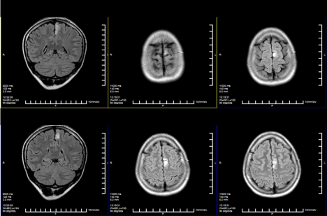
Brain MRI (Figure 1) revealed a focal T2-hyperintensity without mass effect on the mesial face of the left frontal lobe without postcontrast enhancement; spectroscopy findings were consistent with chronic inflammatory lesion (increased choline and reduced N-acetyl- aspartate peaks). Functional MRI revealed reduced activation of the left supplementary motor area, just contiguous to the lesion.

    Figure 1:  Brain MRI (FLAIR sequence) showing a focal cortical lesion in the
left mesial frontal lobe, without mass effect.
CSF oligoclonal bands were detected and there was serum high titre positivity for ANA (>1/640), indicating a possible autoimmune pathogenesis of the disease.
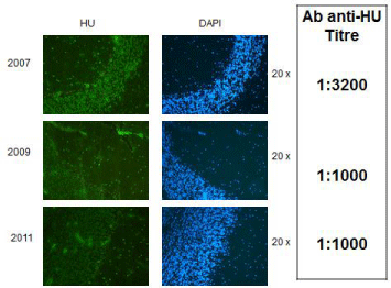
Interestingly, we also found serum positivity for anti-HU antibodies (tested both in IIF and IMMUNOBLOT titre 1/3200, (Figure 2), but repeated complete workup for searching occult neoplasm, tested negative for up to 5 years.
The patient has been treated with oral prednisone (10 mg daily) and the monoclonal antibody rituximab.
Rituximab was administered in May 2007 (375 mg/m² every week for 4 weeks) and repeated once in May 2008 (1000 mg twice 15 days apart) in occasion of recurrence of blood CD19+ cells, tested monthly as marker of rituximab bioactivity, without adverse events.
Stabilisation of the disease was observed (no more recurrence of status epilepticus, slight improvement of paresis and choreoathetotic movements, stabilisation of cognition); the focal brain lesion was stable at subsequent MRI studies and anti-HU antibodies titre decreased at the end of 7-years follow-up (Figure 2). The only remarkable features are sporadic focal motor seizures, well controlled by antiepileptic drugs.

    Figure 2:  Semi-quantitative detection of anti-HU antibodies using
immunofluorescence (Primate Cerebellum, Euroimmun, Luebeck, Germany).
1:100- diluted human serum was incubated with cryosections of primate
cerebellum; bound IgG was visualized using secondary antibodies labeled
with FITC, showing anti-HU antibodies typical fluorescence staining pattern
on cell nuclei in both granular and molecular layers (green). As control, cell
nuclei, stained with DAPI, are shown in blue. Progressive reduction in anti-HU
antibodies titre has been showed during the follow-up.
Magnification 20x. GL: Granular Layer; ML: Molecular Layer; DAPI:
4,6-diamidino-2-phenylindole; FITC: Fluorescein Isothiocyanate.
Discussion
Rasmussen’s encephalitis (Chronic Focal Encephalitis– CFE) is a chronic inflammatory disease of the CNS affecting children characterized by refractory partial seizures with progression to nonconvulsive status epilepticus, cognitive decline, focal cortical lesion and progressive atrophy of the affected hemisphere [1].
The early neuroimaging and neurophysiological features are not characteristic: nevertheless the clinical and pathological characteristics define the diagnosis [1-3].
Recently, limited and atypical forms of CFE have been described, in particular forms of complex partial seizures and choreoathetotic movements of the limbs with focal cortical inflammatory lesions [4].
Non-convulsive refractory status epilepticus has been described in anti-HU associated paraneoplastic disorders and antineuronal antibodies can be present in non-paraneoplastic systemic autoimmune disorders as Sjogren Syndrome or Systemic Lupus Erithematosus (SLE) [5-8].
Our patient showed clinical and radiological characteristics similar to limited forms of CFE with a clear autoimmune background (ANA positive serum, CSF showing oligoclonal bands) in which anti- HU antibodies have been definitely found without neoplasm.
In addition, immunosuppression by the use of Rituximab, a monoclonal antibody with immunosuppressive effect through B-cell depletion used for several autoimmune diseases refractory to conventional therapy, determined stabilisation of the disease and concomitant decrease of anti-HU titre, suggesting their possible pathogenetic role.
Even if not biopsy proven, all these findings suggest the possibility of anti-HU mediated epileptogenic focal encephalitis of the Rasmussen’s type: according to experimental data anti-HU antibodies are directed against RNA structures and impaired the synthesis of proteins leading to widespread functional deficits as shown by the plethora of manifestations of anti-HU associated paraneoplastic neurological disorders, including limbic encephalitis, movement disorders, ataxia, progressive dementia and seizures [9-11].
In conclusion, we suggest the research of anti-neuronal antibodies in every case of unsolved epileptogenic encephalitis in particular those occurring with non-convulsive status epilepticus, and the use of rituximab in the treatment of atypical refractory epilepsy in which an autoimmune pathogenesis can be supported by the laboratory findings, as also suggested by an ongoing pilot trial of the use of rituximab in CFE [12,13].
References
- Varadkar S, Bien CG, Kruse CA, Jensen FE, Bauer J, Pardo CA, et al. Rasmussen's encephalitis: clinical features, pathobiology, and treatment advances. Lancet Neurol. 2014; 13: 195-205.
- Türkdogan-Sözüer D, Ozek MM, Sav A, Dinçer A, Pamir MN. Serial MRI and MRS studies with unusual findings in Rasmussen's encephalitis. Eur Radiol. 2000; 10: 962-966.
- Freeman JM. Rasmussen's syndrome: progressive autoimmune multi-focal encephalopathy. Pediatr Neurol. 2005; 32: 295-299.
- Gambardella A, Andermann F, Shorvon S, Le Piane E, Aguglia U. Limited chronic focal encephalitis: another variant of Rasmussen syndrome? Neurology. 2008; 70: 374-377.
- Nahab F, Heller A, Laroche SM. Focal cortical resection for complex partial status epilepticus due to a paraneoplastic encephalitis. Neurologist. 2008; 14: 56-59.
- Greenlee JE. Anti-Hu antibody and refractory nonconvulsive status epilepticus. J Neurol Sci. 2006; 246: 1-3.
- Benyahia B, Amoura Z, Rousseau A, Le Clanche C, Carpentier A, Piette JC, et al. Paraneoplastic antineuronal antibodies in patients with systemic autoimmune diseases. J Neurooncol. 2003; 62: 349-351.
- Kano O, Arasaki K, Ikeda K, Aoyagi J, Shiraishi H, Motomura M, et al. Limbic encephalitis associated with systemic lupus erythematosus. Lupus. 2009; 18: 1316-1319.
- Carpentier AF, Chassande B, Amoura Z, Benyahia B, Piette JC, Delattre JY. Systemic lupus erythematosus with anti-Hu antibodies and polyradiculoneuropathy. Neurology. 2001; 57: 558-559.
- Totland C, Aarseth J, Vedeler C. Hu and Yo antibodies have heterogeneous avidity. J Neuroimmunol. 2007; 185: 162-167.
- Voltz RD, Posner JB, Dalmau J, Graus F. Paraneoplastic encephalomyelitis: an update of the effects of the anti-Hu immune response on the nervous system and tumour. J Neurol Neurosurg Psychiatry. 1997; 63: 133-136.
- Granata T, Fusco L, Gobbi G, Freri E, Ragona F, Broggi G, et al. Experience with immunomodulatory treatments in Rasmussen's encephalitis. Neurology. 2003; 61: 1807-1810.
- A Pilot Study of the Use of Rituximab in the Treatment of Chronic Focal Encephalitis (Identifier NCT00259805).
Citation: Capobianco M, Sperli F, Malentacchi M, Valentino P, Sala A, et al. Anti-HU Antibodies Associated Focal Epileptogenic Encephalitis Successfully Treated with Rituximab: A New Limited form of Chronic Focal Encephalitis of the Rasmussen’s Type?. Austin J Clin Neurol 2015;2(5): 1043. ISSN : 2381-9154