Case Report
Austin J Clin Neurol 2015;2(5): 1046.
Reversible Cerebral Vasoconstriction Syndrome with Atypical Subarachnoid Hemorrhage: A First Case Report
Gentile M¹*, De Vito A², Groppo E¹, Tola MR² and Azzini C²
¹Department of Biomedical Sciences and Medical Surgical Sciences, University of Ferrara, Italy
²Department of Neurology, Stroke Unit, Arcispedale S. Anna Ferrara, Italy
*Corresponding author: Mauro Gentile, Department of Biomedical Sciences and Medical Surgical Sciences, University of Ferrara, via Aldo Moro 8, 44123 Ferrara, Italy
Received: April 02, 2015; Accepted: May 05, 2015; Published: May 15, 2015
Abstract
Reversible cerebral vasoconstriction syndrome represents a group of syndromes characterised by prolonged, reversible, segmental vasoconstriction of the cerebral arteries, usually associated with acute, severe, “thunderclap” headache with or without additional focal neurological deficits and seizures. The major complications of this syndrome are localised cortical subarachnoid haemorrhages and ischaemic or haemorrhagic strokes. Subarachnoidhemorrhage represents the most frequent hemorrhagic complication and it is often mild, focal and superficial and involves few high sulci in the convexity. To our knowledge this report represents the first case of atypical subarachnoid hemorrhage localised in the basal cistern associated with reversible cerebral vasoconstriction syndrome.
We report a 52 year old woman presented severe sudden onset occipital headache episodes. The neurological examination revealed only horizontal nystagmus in left sight direction; vital signs were normal. Neuroimaging scans (CT and MRI) showed a mild diffuse cerebral swelling without focal lesions. The following days she a thunderclap type headache recurred acutely and a new brain CT showed subarachnoid hemorrhage in the basal cisterns. Non invasive angiography demonstrated a wide segmental vasoconstriction of the cerebral arteries with dilatations of the post-stenotic segments suggesting reversible cerebral vasoconstriction syndrome. New non invasive angiography obtained after 3 months showed a complete resolution of the previous segmental arterial vasoconstrictions.
Keywords: Reversible cerebral vasoconstriction syndrome; Call-Fleming syndrome; subarachnoid hemorrhage
Introduction
Reversible cerebral vasoconstriction syndrome (RCVS) was first recognized by Call and Fleming in 1988 [1]. RCVS represents a group of syndromes characterized by prolonged, reversible, segmental vasoconstriction of the cerebral arteries, usually associated with acute, severe, “thunderclap” headache with or without additional focal neurological deficits and seizures. It usually resolves in 1-3 months after onset [2].
The disease pathophysiological mechanism is still unknown. However it is possible to recognize a specific cause of RCVS only in 25- 60% of patients as postpartum vasospasm (with or without eclampsia or pre-eclampsia), exposure to vasoactive or immunosuppressant drugs, exposure to blood products, catecholamine secreting tumors, hypercalcemia, porphyria, head trauma, neurosurgery, subdural spinal haematoma, carotid endoarterectomy, cerebral venous thrombosis, CSF hypotension, autonomic dysreflexia, phenytoin intoxication [3], hormonal ovarian stimulation for intrauterine insemination [4], LES [5], tonsillectomy [6], high altitude [7]. Therefore there is still a high percentage of idiopathic cases.
The incidence of this condition is probably underestimated, particularly because of the undiagnosed benign forms without focal neurological deficits or seizures. Females seem to be more affected than males with a 2-10:1 ratio [2,8,9].
The RCVS major complications are localised cortical subarachnoid haemorrhages (SAH) and ischaemic or haemorrhagic strokes [8,10- 14]. The former represents the most frequent RCVS hemorrhagic manifestation [14].
We report a case of RCVS associated with atypical SAH.
Case Presentation
A 52 year old woman was admitted to our Emergency Department because of severe sudden onset occipital headache episodes associated to asthenia in the last ten days. She had a past history of short transient blurred vision episodes occurring in her half visual field (especially the left one) without any other associated symptoms. She did not use any drug.
At admission to our Neurologic Unit the examination revealed only horizontal nystagmus in left gaze. Vital signs, including blood pressure (120/80 mmHg), were normal. Brain computed tomography (CT) scan and magnetic resonance imaging (MRI) showed a mild diffuse cerebral swelling without focal lesions.
In the evening of day 1, while headache was improving, she developed spatial disorientation and mild speech slowdown without other neurological deficits. EEG registration revealed diffuse slow abnormalities with epileptiform focal aspects in the left temporal region.
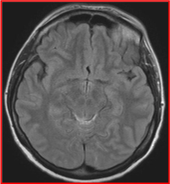
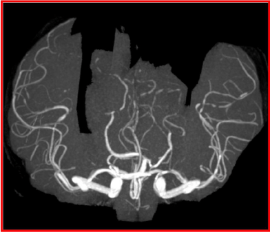
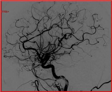
The following day a thunderclap type headache recurred acutely with pulsating severe occipital pain and mild neck stiffness. To rule out a SAH, a new cerebral CT was performed showing SAH in the basal cisterns with diffuse cerebral swelling. MRI with venous sequences confirmed SAH and excluded cerebral sinuses thrombosis (Figure 1). Non invasive angiography (MR angiography) and invasive angiography demonstrated a wide segmental vasoconstriction of the cerebral arteries with dilatations of the post-stenotic segments (“string of beads” appearance), without aneurysms or other vascular malformations (Figure 2,3).

Figure 1: Magnetic resonance with evidence of SAH in the basal cistern.

    Figure 2:  Non invasive and invasive angiography show a wide segmental
vasoconstriction of the cerebral arteries with dilatations of the post-stenotic
segments (“string of beads” appearance), without aneurysms or other
vascular malformations.

    Figure 3:  Non invasive and invasive angiography show a wide segmental
vasoconstriction of the cerebral arteries with dilatations of the post-stenotic
segments (“string of beads” appearance), without aneurysms or other
vascular malformations.
Transcranial sonography was normal, it did not show flow accelerations of the intracranial arteries. Complete blood count, biochemistry and coagulation were normal as well as ESR, CRP, autoimmunity screening and serological tests. Furthermore the CSF analysis was normal.
In the following days the patient progressively improved; after 2 weeks she was asymptomatic, neurological examination was normal and she was discharged at home.
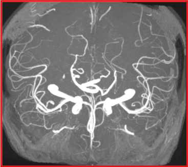
An MR angiography obtained after 3 months showed a complete resolution of the previous segmental arterial vasoconstrictions (Figure 4).

    Figure 4:  3 month’s follow-up non invasive angiography show complete
resolution of the previous segmental vasoconstriction.
No further episodes of “thunderclap” headache occurred during the 4-years follow-up period and her neurological examination is still negative thenceforth.
Discussion
RCVS is characterized by prolonged, reversible, segmental vasoconstriction of the cerebral arteries. The first symptom is usually “thunderclap” headache [15], with a characteristic explosive onset. Often there is more than one “thunderclap” headache episode as in our case [9]. Severe headache episodes can be the only RCVS manifestations or they can be associated to other neurological symptoms [8]. Sometimes it’s possible to identify a trigger as Valsalva manoeuvre, emotional or physical stress, sudden head movements, but commonly onset can be spontaneous as in our patient. Other concomitant or subsequent neurological manifestations are represented by transient or persistent focal deficits, including visual, sensory or speech disturbances, hemiparesis, ataxia and seizures [2,8,9]. Our patient developed a transient visuo-spatial disorientation. Moreover in the patient’s past history there were some episodes characterized by transient blurred vision in half visual field possibly related to former reversible vasospastic events.
As in our report, laboratory tests are usually not significant.
The main RCVS complications are intracranial haemorrhage or brain infarction. In the available literature a wide range of haemorrhagic or ischemic complications are reported because of patient’s series heterogeneity and small patient’s number. The greatest case series comprises 139 patients [9]: 81% developed brain lesions including brain infarction (39%), SAH (34%), parenchymal hemorrhage (20%) and brain oedema (38%). In some patients authors described more than one complication. SAH represents the most frequent hemorrhagic complication and it is often mild, focal, superficial and involves few high sulci in the convexity [9,11,14,16] while in our report CT and MRI scan revealed a hemorrhagic lesion in the basal cisterns. To our knowledge this is the first description of non convexity SAH associated with RCVS. Subsequent transfemoral angiography confirmed a typical RCVS artery pattern and excluded the presence of vascular malformations.
RCVS diagnosis requires the evidence of a typical angiographic pattern characterised by a bilateral multiple “string of beads” appearance of the cerebral arteries caused by segmental vasoconstrictions and post-stenotic dilatations, revealed by noninvasive or invasive angiography. Extracranial segments of cerebral afferent arteries are rarely affected [9]. According to the diagnostic criteria proposed by Calabrese et al. [2] vasospasm reversibility must be demonstrated by an angiographic control within 12 weeks after onset. In our patient we performed a non-invasive MR angiographic study that showed complete resolution of the previous vasospasm after 3 months.
Transcranial Doppler ultrasonography can be useful to monitor vasospasm avoiding more invasive and expensive tests during followup [17,18]; our patient had a normal intracranial arteries Doppler velocity spectrum. Usually outcome is good with a 0-1 score at modified Rankin Scale as described in about 80% of patients [9]; however it strictly depends on complications occurrence. Our patient is symptoms-free after a 4 years follow up.
Major differential diagnoses have to be made with primary angiitis of CNS (PACNS) but in the latter headache course is more often subacute or chronic. Other remarkable findings in PACNS are the abnormal CFS analysis and the vasospasm irreversibility. Our patient showed a normal CSF analysis and vasospasm reversibility. In order to differentiate RCVS from PACNS high resolution contrastenhanced vessel wall MRI has been recently proposed because this technique shows arterial wall enhancement typical of PACNS patients [19].
In conclusion to our knowledge this is the first report of a patient affected by RCVS associated with atypical SAH localization (basal cisterns).
References
- Call GK, Fleming MC, Sealfon S, Levine H, Kistler JP, Fisher CM. Reversible cerebral segmental vasoconstriction. Stroke. 1988; 19: 1159-1170.
- Calabrese LH, Dodick DW, Schwedt TJ, Singhal AB. Narrative review: reversible cerebral vasoconstriction syndromes. Ann Intern Med. 2007; 146: 34-44.
- Ducros A. Reversible cerebral vasoconstriction syndrome. Lancet Neurol. 2012; 11: 906-917.
- Freilinger T, Schmidt C, Duering M, Linn J, Straube A, Peters N. Reversible cerebral vasoconstriction syndrome associated with hormone therapy for intrauterine insemination. Cephalalgia. 2010; 30: 1127-1132.
- Ashraf VV, Bhasi R, Ramakrishnan KG, Praveenkumar R, Girija AS. Reversible cerebral vasoconstriction syndrome in a patient with systemic lupus erythematosus. Neurol India. 2012; 60: 635-637.
- Trinidade A, Shakeel M, Majithia A, Solbach T, Dhillon RS. Call-fleming syndrome: an extraordinary complication of tonsillectomy. J Coll Physicians Surg Pak. 2010; 20: 822-824.
- Neil WP, Dechant V, Urtecho J. Pearls & oy-sters: reversible cerebral vasoconstriction syndrome precipitated by ascent to high altitude. Neurology. 2011; 76: e7-9.
- Ducros A, Boukobza M, Porcher R, Sarov M, Valade D, Bousser MG. The clinical and radiological spectrum of reversible cerebral vasoconstriction syndrome. A prospective series of 67 patients. Brain. 2007; 130: 3091-3101.
- Singhal AB, Hajj-Ali RA, Topcuoglu MA, Fok J, Bena J, Yang D, et al. Reversible cerebral vasoconstriction syndromes: analysis of 139 cases. Arch Neurol. 2011; 68: 1005-1012.
- Chen SP, Fuh JL, Lirng JF, Chang FC, Wang SJ. Recurrent primary thunderclap headache and benign CNS angiopathy: spectra of the same disorder? Neurology. 2006; 67: 2164-2169.
- Edlow BL, Kasner SE, Hurst RW. Reversible cerebral vasoconstriction syndrome associated with subarachnoid hemorrhage. Neurocrit Care. 2007; 7: 203-210
- Moskowitz SI, Calabrese LH, Weil RJ. Benign angiopathy of the central nervous system presenting with intracerebral hemorrhage. Surg Neurol. 2007; 67: 522-527.
- Ducros A, Bousser MG. Reversible cerebral vasoconstriction syndrome. Pract Neurol. 2009; 9: 256-267.
- Ducros A, Fiedler U, Porcher R, Boukobza M, Stapf C, Bousser MG. Hemorrhagic manifestations of reversible cerebral vasoconstriction syndrome: frequency, features, and risk factors. Stroke. 2010; 41: 2505-2511.
- Headache Classification Subcommittee of the International Headache Society. The International Classification of Headache Disorders: 2nd edtn. Cephalalgia. 2004; 24: 9-160.
- Ansari SA, Rath TJ, Gandhi D. Reversible cerebral vasoconstriction syndromes presenting with subarachnoid hemorrhage: a case series. J Neurointerv Surg. 2011; 3: 272-278.
- Bogousslavsky J, Despland PA, Regli F, Dubuis PY. Postpartum cerebral angiopathy: reversible vasoconstriction assessed by transcranial Doppler ultrasounds. Eur Neurol. 1989; 29: 102-105.
- Chen SP, Fuh JL, Chang FC, Lirng JF, Shia BC, Wang SJ. Transcranial color doppler study for reversible cerebral vasoconstriction syndromes. Ann Neurol. 2008; 63: 751-757.
- Mandell DM, Matouk CC, Farb RI. Vessel wall MRI to differentiate between reversible cerebral vasoconstriction syndrome and central nervous system vasculitis: preliminary results. Stroke. 2012; 43: 860-862.