Case Report
Austin J Clin Neurol 2015;2(7): 1061.
Painful Leg in a Toddler: Consider Idiopathic Neuralgic Amyotrophy
Kliffen J7¹*, Niermeijer JMF² and Engelen M²
¹Department of Neurology, Sint Lucas Andreas Hospital, Netherlands
²Department of Pediatric Neurology, Academic Medical Center Amsterdam, Netherlands
*Corresponding author: Kliffen J, Department of Neurology Sint Lucas Andreas Hospital, PO Box 9243, 1006 AE, Amsterdam, Netherlands
Received: May 06, 2015; Accepted: June 21, 2015; Published: June 30, 2015
Abstract
Idiopathic neuralgic amyotrophy (INA) is a plexopathy with acute onset of severe pain followed by loss of muscle strength and atrophy of the muscles innervated by the involved plexus. Involvement of the brachial plexus is characteristic but occasionally the lumbosacral plexus is involved. This is denominated as ‘idiopathic lumbosacral plexus neuropathy’. INA can occur at any age. This case illustrates the diagnostic course in a toddler with an ‘idiopathic lumbosacral plexus neuropathy’. Even as a recognizable clinical picture, INA remains a diagnosis by exclusion.
Keywords: Neuralgic amyotrophy; Pediatric; Plexopathy; Lumbosacral plexus
Abbreviations
INA: Idiopathic Neuralgic Amyotrophy; CSF: Cerebrospinal Fluid; HNA: Hereditary Neuralgic Amyotrophy
Background
Acute onset of severe pain in a limb, followed by loss of muscle strength and muscle atrophy gives suspicion of idiopathic neuralgic amyotrophy (INA). Our case illustrates that INA also involves the lumbosacral plexus and that it occurs at young age as well. This makes that INA is part of the differential diagnosis in a child with a painful lower limb. There are demonstrable clues towards the diagnosis.
Case Presentation
A 1 year and 8 months old girl was brought in the outpatient department with a painful right lower limb present since three weeks. The pain made it difficult to sleep at night. There was no previous history of trauma or infection. Developmental milestones and further medical history were unremarkable.
Neurological evaluation showed that she preferred to stand on the left leg. Her right foot was in an exorotation position on ambulation. Manipulation of the leg was painful. There was a flaccid paresis and atrophy of the proximal and distal muscles of the right leg. Patellar tendon reflex and ankle jerk were absent on the right leg. Sensory function could not be examined properly. She was also analyzed by an orthopedic surgeon, no other orthopedic abnormalities were found.
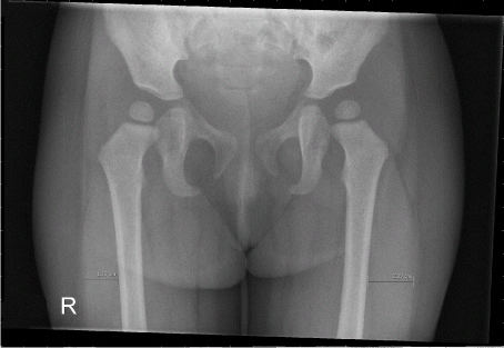
At the initial presentation symptoms and signs were interpreted as most compatible with a lumbosacral plexopathy. The muscle atrophy of the right leg was even visible on the X-ray of the pelvic area (Figure). To exclude treatable causes of plexopathy like inflammation, infection or space occupying lesions further investigations were done. Blood tests and MRI of the spine and lumbosacral plexus were unremarkable. Cerebrospinal fluid (CSF) cell count, protein, and glucose levels were normal. Limited electrophysiological studies, only nerve conduction velocity (the child was uncooperative), did not show any abnormalities.

    Figure 1:  Plain X-ray of the pelvic area. The vastusexternus of the quadriceps
femoris muscle on the right side measures 1,58 cm compared to 2,00 cm on
the left side.
At follow-up visit, 10 weeks after onset of the complaints, there was clear improvement. Proximal atrophy of the right leg was still visible but ambulation was normal. Tendon reflexes were restored to a normal symmetrical patron. The diagnosis of idiopathic neuralgic amyotrophy (INA) was considered knowing that this disease does not exclusively present with a neuropathy of brachial plexus. The natural course is consistent with the diagnosis.
Discussion
INA is a plexopathy with acute onset of severe pain followed by loss of muscle strength and atrophy of the muscles innervated by the involved part of the plexus. The exact incidence of INA is not known. INA can present at any age, ranging from the neonatal period till geriatric age [1]. Pain is less often a presenting symptom of INA in children, making it more difficult to recognize [1]. Our patient did present with pain but the pain was solely located in the lower limb. It is less known that INA can also involve the lumbosacral plexus, in which case the disorder is called ‘idiopathic lumbosacral plexus neuropathy’ [2]. Involvement of the lumbosacral plexus in INA patients is less prevalent. It is estimated that less than ten percent of INA patients have involvement of the lower limb [3]. However this is more often than involvement of other peripheral nerves like the phrenic nerve, recurrent laryngeal nerve or peripheral autonomic nerves [3].
Even as a recognizable clinical picture, INA remains a diagnosis by exclusion. Lumbosacral plexopathies have a broad range of causes. The most important causes are vascular (proximal diabetic neuropathy), infectious (neuroborreliosis or HIV infection), spaceoccupying lesion (for example neoplastic invasion or retroperitoneal hematoma) and trauma. The diagnostic evaluation includes laboratory investigations, electrophysiological studies (nerve conduction and myography) and neuroimaging (MRI). Further, CSF examination can be necessary to exclude an occult neuroinfection or neoplasia if suspected. In approximately ten percent of INA patients CSF analysis shows an elevated protein level which is a nonspecific fact [3]. Nerve conduction studies can confirm an axonal plexopathy, and exclude a monoradiculopathy, pressure neuropathy or demyelinisation. An important pitfall with myography is that it can be normal if the affected muscles are not tested. On top of this electrophysiological studies can be a challenge in young children [4]. In a minority of patients MRI scan of the plexus shows focal T2 hyperintensity or focal thickening of the plexus [3].
INA is considered an auto-immune disease. In support of this hypothesis, attacks are often preceded by activation of the immune system, for instance infection, operation or vaccination. Physical stress is identified as a risk factor [3]. The conception is that mechanical disturbance leads to disruption of the blood nerve barrier which gives opportunity for attack to the peripheral nerves [3].
Besides idiopathic neuralgic amyotrophy, a hereditary form exist which is less common then INA. In hereditary neuralgic amyotrophy (HNA) neuralgia attacks will recur more often. Thus family history is a relevant part of history taking and discussing the prognosis in patients with INA. HNA has an autosomal dominant inheritance pattern. In some affected families the associated point mutation or duplication in the SEPT9 gene on chromosome 17 can be found [5]. Typical dysmorphic features are seen often in these families like small, round faces, hypotelorism and short stature [6]. More than half of patients with HNA have involvement of nerves besides the brachial plexus [3]. On average the first appearance of HNA is earlier in life compared to INA [3].
Given the hypothesis that INA is an immunologically mediated syndrome, treatment with immunosuppressive medication would be rational. Indeed oral prednisolone seems effective for pain relief and strength recovery in observational studies in the acute phase of INA [7,8]. This effect needs to be verified in a prospective, randomized trial. Symptomatic treatment needs to be adapted to the stage and type of pain. Usual care comprises treatment of the acute and often severe (neuropathic) pain and guidance for supportive care. Prevention of contractures is of uttermost importance. In later stages residual neuropathic and mechanical hypersensitivity pain can remain. Paretic muscles and compensatory mechanisms can lead to musculoskeletal pain and overburden is a hazard. Unfortunately recovery for children is less favorable, less than half of patients gain full recovery [1]. However the average follow-up time in this evaluation was relatively short (10 months). In adults it is known that recovery time can extend for over more than two years.
References
- van Alfen N, Schuuring J, van Engelen BG, Rotteveel JJ, Gabreëls FJ. Idiopathic neuralgic amyotrophy in children. A distinct phenotype compared to the adult form. Neuropediatrics. 2000; 31: 328-332.
- van Alfen N, van Engelen BG. Lumbosacral plexus neuropathy: a case report and review of the literature. Clin Neurol Neurosurg. 1997; 99: 138-141.
- van Alfen N, van Engelen BG. The clinical spectrum of neuralgic amyotrophy in 246 cases. Brain. 2006; 129: 438-450.
- Pitt MC. Nerve conduction studies and needle EMG in very small children. Eur J Paediatr Neurol. 2012; 16: 285-291.
- van Alfen N. Clinical and pathophysiological concepts of neuralgic amyotrophy. Nat Rev Neurol. 2011; 7: 315-322.
- Laccone F, Hannibal MC, Neesen J, Grisold W, Chance PF, Rehder H. Dysmorphic syndrome of hereditary neuralgic amyotrophy associated with a SEPT9 gene mutation--a family study. Clin Genet. 2008; 74: 279-283.
- van Eijk JJ, van Alfen N, Berrevoets M, van der Wilt GJ, Pillen S, van Engelen BG. Evaluation of prednisolone treatment in the acute phase of neuralgic amyotrophy: an observational study. J Neurol Neurosurg Psychiatry. 2009; 80: 1120-1124.
- van Alfen N, van Engelen BG, Hughes RA. Treatment for idiopathic and hereditary neuralgic amyotrophy (brachial neuritis). Cochrane Database Syst Rev. 2009; : CD006976.