Case Report
Austin J Clin Neurol 2016; 3(1): 1089.
Delayed Onset of Contralateral Abducens Nerve Palsy after Embolization of Carotid Cavernous Sinus Fistula: A Case Report
Yao CA¹, Tsou HK², Sheehan J² and Pan HC1,3*
¹Department of Neurosurgery, Taichung Veterans General Hospital, Taiwan
²Department of Neurosurgery, University of Virginia, USA
³Faculty of Medicine, School of Medicine, National Yang- Ming University, Taiwan
*Corresponding author: Hung-Chuan Pan, Department of Neurosurgery, Taichung Veterans General Hospital, 1650 Taiwan, Boulevard Sec.4, 40705 Taichung, Taiwan
Faculty of Medicine, School of Medicine, National Yang- Ming University, Taipei, Taiwan
Received: July 21, 2016; Accepted: August 27, 2016; Published: August 30, 2016
Abstract
A 28 year old female harbored a left sided direct carotid cavernous sinus fistula. An attempt with coil embolization proved unsuccessful, and the carotid cavernous sinus fistula was then successfully trapped. Following the intervention, the patient had the improvement with her chemosis and protruding status over left eye; there were no neurological deficits associated with the procedure. However, ten years later, the patient complained about new onset of diplopia. Neurological examination revealed right abducens nerve palsy. Magnetic resonance angiography showed the engorgement of the right side cavernous sinus and increased diameter of the right internal carotid artery, but there was no definite tumor or aneurysm adjacent to cavernous sinus or skull base. The delayed onset of contralateral abducens nerve palsy is rare. We reported this rare case and conducted a literature review of similar cases.
Keywords: Carotid cavernous sinus fistula; Abducens nerve palsy; Embolization
Introduction
In embolization of a direct carotid cavernous fistula (CCF), the reported complication rates vary from 1% to 40%, and complications include an ocular nerve palsy and carotid artery occlusion [1,2]. The ophthalmoplegia may be a complication of coil embolization from mass effect of a progressive thrombosis within the cavernous sinus or direct traumatic damage from the coils or microcatheters themselves [3]. In some occasions, the ophthmalmoplegia was proposed to be muscle hypoxia rather than cranial nerve palsy because of incomplete or uneven recovery even with total occlusion of the fistula [4,5].
The increased blood flow contralateral to the occlusion site was reported in patients subjected to occlusion of internal carotid artery disease [6]. The incidence of late consequence of internal carotid ligation for the treatment of cavernous sinus aneurysm was 24% and complications included transient ischemia attack, subarachnoid hemorrhage (SAH), or fatal death [7]. To date, there was no report of delayed onset of ophthalmoplegia contralateral to the location of CCF after embolization. In this report, we present a case of contralateral abducens nerve palsy after the obliteration of a direct CCF by the total occlusion of the internal carotid artery. We also have conducted a literature review for similar cases.
Case Presentation
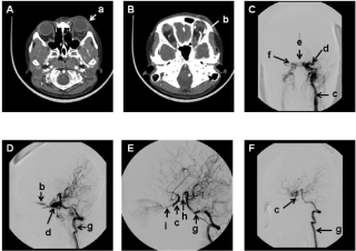
A 28 year old female suffered a motorcycle accident, and she developed chemosis and progressive protrusion over left eye on July 1, 2005 (Figure 1A). The computed tomography angiography (CTA) showed enlargement of left superior ophthalmic veins (Figure 1B). Cerebral angiography showed a direct CCF from left internal carotid artery (ICA) (Figure 1C) and lateral vertebral artery angiography further confirmed left direct CCF with temporal occlusion of the left ICA (Figure 1D). The attempting to embolize the fistula with Guglielmi detachable coils from left ICA approach failed. The temporal occlusion of the left ICA with a balloon test was conducted, and the patient tolerated the test occlusion without neurological deterioration. The patient was subjected to the trapping of the left ICA. The post embolization vertebral angiography in anteriorposterior and lateral projection showed the total obliteration of the CCF (Figure 1E and F). The patient demonstrated substantial clinical improvement with resolution of her chemosis and protruding eye.

    Figure 1: Representative imaging findings in this patient 3 months after the traffic accident and then following embolization (A) left eye ball protruding in axial CT
scan 3 months after the traffic accident (B) Engorgement of the left ophthalmic veins in CTA (C) Left carotid cerebral angiography in posterior-anterior projection (D)
Vertebral angiography in lateral view after balloon occlusion of the left ICA (E ) Post embolization of vertebral cerebral angiography in posterior-anterior projection
(F) Post embolization with vertebral cerebral angiography in lateral view. a:left protruding eye; b: left superior ophthalmic vein; c: left internal carotid artery; d: left
cavernous sinus; e: intercavernous connecting veins; f:right cavernous sinus; g: left vertebral artery; h:posterior communicating artery; i: ophthalmic artery.
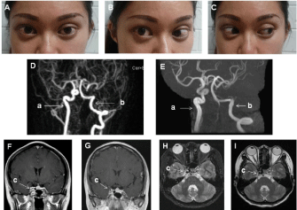
Approximately 10 years after the left sided CCF trapping, the patient started to experiencing diplopia, but there was no definite EOM limitation at that time. Ten months later, the neurological examination only showed a limitation of lateral gaze play over right eye in favor of definitive right abducens nerve palsy (Figure 2A,B and C). Magnetic resonance angiography (MRA) revealed the increased size of the right ICA (4.9 mm in diameter) as compared to the imaging obtained 3 months after embolization (4.1 mm in diameter) (Figure 2D and E). Magnetic resonance imaging (MRI ) also showed an increased size of the cavernous sinus demonstrated in coronal (14.6x9.1mm) and axial (14.5 mm) view as compared to comparable neuro-images obtained 3 months after the embolization (13.5 x 8.7 mm, 13.5 mm, respectively) (Figure 2F,G,H and I). In addition, there was no definite tumor or aneurysm adjacent to cavernous sinus or skull base in MRI examination. The patient remains with a right abducens nerve palsy and has shown no improvement in recent outpatient clinic visits.

    Figure 2: Representative right abducens nerve palsy and associated MRI imaging findings (A) both eyes in central position (B) both eyes turned to right (C) both
eyes turned to left (D) MRA of the brain 3 months after embolization (E) MRA of brain 10 years after embolization (F) T1 weighted MRI with contrast administration
in coronal views 3 months after embolization (G) T1 weighted MRI with contrast administration in coronal views 10 years after embolization (H) T2 weighted MRI
3 months after embolization in axial view (I) T2 weighted MRI 10 years after embolization in axial view. a: right internal carotid artery; b: left vertebral artery; c:
cavernous sinus.
Discussion
The treatment of a direct carotid cavernous fistula included transarterial and trans-venous approach by using balloon, coil, or stent, is intended to not only to obliterate the fistula but also to preserve the patency of ICA [8-11]. In some occasions, the technique to obliterate the fistula without compromising the ICA patency is difficult. The trapping of ICA without disruption of efficacy of collateral circulation is generally a reasonable treatment option [12]. In this case report, the attempting to embolize the fistula failed and then the procedure was converted to trapping the ICA. Following the procedure, the patient gained improved clinically without any immediate complications.
The treatment complications for a direct CCF consist of vessel injury, mass effect from the coil or balloon, and alterations in the drainage of the venous or arterial systems. The symptoms include the immediate occurrence of subarachnoid hemorrhage, intracerebral hemorrhage, ophthalmoplegia, and brain infarction and late complication of fistula recurrence or ipsilateral ophthalmoplegia [1- 5,13]. In this case report, the patient was selected for trapping ICA to obtain immediate symptomatic relief. However, delayed onset of contra-lateral abducens nerve palsy was noted 10 years later. This complication does not appear to be a direct but rather an indirect result of the C-C fistula embolization.
Complications related to ICA occlusion in direct CCF treatment are rare. There were some reports detailing the late onset of an indirect CCF from ICA occlusion but these cases did not report the occurrence of ophthalmoplegia [12-17]. The literature associated with cavernous sinus aneurysms treated by the ICA occlusion reflects similar hemodynamic changes as with CCF obliteration by ICA trapping. According to the report of cavernous sinus aneurysm treated by ICA ligation, there were as high as 25% of patients who presented with brain ischemia or SAH without influencing the function of extra-ocular nerves [7]. In the current case, a left CCF was occluded by left ICA occlusion, but the right abducens nerve palsy reflects less of a direct effect from left ICA occlusion.
The hemodynamic alteration in stroke patients in total or partial occlusion of the ICA show decreased blood flow in on the occluded side but an increase of the flow of contralateral side [6,7]. In the current patient, the left ICA was occluded with compensated engorgement of the right ICA. In addition, the tortuous morphology of the ICA was noted in right cavernous sinus portion which may exert high pressure on neural structures within the cavernous sinus. Furthermore, there was no definite tumor over cavernous sinus or skull base demonstrated in MRI examination. This phenomenon could explain the delayed right abducens nerve palsy contralateral to left ICA occlusion after CCF embolization.
Conclusion
The delayed onset of contralateral abducens nerve palsy was less likely a direct effect of the embolization of a CC fistula. The hemodynamic alteration with increased blood flow contralateral to the occluded CC fistula site may contribute to delayed onset cranial nerve dysfunction.
Acknowledgement
We thank you for Mr. Liang-Yi Pan for preparation of the photography in this manuscript.
References
- Gupta AK, Purkayastha S, Krishnamoorthy T, Bodhey NK, Kapilamoorthy TR, Kesavadas C, et al. Endovascular treatment of direct carotid cavernous fistulae: a pictorial review. Neuroradiology. 2006; 48: 831-839.
- Gemmete JJ, Ansari SA, Gandhi DM. Endovascular techniques for treatment of carotid-cavernous fistula. J Neuroophthalmol. 2009; 29: 62-71.
- Yoshida K, Melake M, Oishi H, Yamamoto M, Arai H. Transvenous embolization of dural carotid cavernous fistulas: a series of 44 consecutive patients. AJNR Am J Neuroradiol. 2010; 31: 651-655.
- Das S, Bendok BR, Novakovic RL, Parkinson RJ, Rosengart AJ, Macdonald RL,et al. Return of vision after transarterial coiling of a carotid cavernous sinus fistula: case report. Surg Neurol. 2006; 66: 82-85.
- Bink A, Berkefeld J, Lüchtenberg M, Gerlach R, Neumann-Haefelin T, Zanella F, et al. Coil embolization of cavernous sinus in patients with direct and dural arteriovenous fistula. Eur Radiol. 2009; 19: 1443-1449.
- Schneider PA, Rossman ME, Bernstein EF, Torem S, Ringelstein EB, Otis SM. Effect of internal artery occlusion on intracranial hemodynamics transcranial Doppler evaluation and clinical correlation. Stroke. 1988: 19: 589-593.
- Roski RA, Spetzler RF, Nulsen FE. Late complications of carotid ligation in the treatment of intracranial aneurysms. J Neurosurg. 1981; 54: 583-587.
- Ng PP, Higashida RT, Cullen S, Malek R, Halbach VV, Dowd CF. Endovascular strategies for carotid cavernous and intracerebral dural arteriovenous fistulas. Journal of Neurosurgery. 2003; 15: 1-6.
- Lewis AI, Tomsick TA, Tew JM Jr, Lawless MA. Long-term results in direct carotid-cavernous fistulas after treatment with detachable balloons. J Neurosurg. 1996; 84: 400-404.
- Halbach VV, Higashida RT, Hieshima GB, Hardin CW, Yang PJ. Transvenous embolization of direct carotid cavernous fistulas. AJNR Am J Neuroradiol. 1988; 9: 741-747.
- Gomez F, Escobar W, Gomez AM, Gomez JF, Anaya CA. Treatment of carotid cavernous fistulas using covered stents: midterm results in seven patients. AJNR Am J Neuroradiol. 2007; 28: 1762-1768.
- Coley SC, Pandya H, Hodgson TJ, Jeffree MA, Deasy NP. Endovascular trapping of traumatic carotid-cavernous fistulae. AJNR Am J Neuroradiol. 2003; 24: 1785-1788.
- Teng MM, Chang T, Pan DH, Chang CN, Huang CI, Guo WY, et al. Brainstem edema: an unusual complication of carotid cavernous fistula. AJNR Am J Neuroradiol. 1991; 12: 139-142.
- Serbinenko FA. Balloon catheterization and occlusion of major cerebral vessels. J Neurosurg. 1974; 41: 125-145.
- Mathis JM, Barr JD, Jungreis CA, Yonas H, Sekhar LN, Vincent D. Temporary balloon test occlusion of the internal carotid artery: Experienced in 500 cases. AJNR Am J Neuroradiol. 1995; 16: 749-754.
- van der Schaaf IC, Brilstra EH, Buskens E, Rinkel GJ. Endovascular treatment of aneurysm in the cavernous sinus: A systematic review on balloon occlusion of the parent vessels and embolization with colis. Stroke. 2002; 33: 313-318.
- Dang NV, Hai NT, Cuong TC. Recurrent carotico-cavernous fistula after internal carotid artery ligation: a case with embolization of the fistula via contralateral internal carotid artery approach. Interv Neruoradiol. 2014; 20: 482-486.