Case Report
Austin J Clin Neurol 2017; 4(1): 1098.
Bilateral Caudate Head Infarct Following Olfactory Groove Meningioma Resection
Desai K*, Spiegel LL, Godfrey RR, Damani R and Bershad EM
Department of Neurology & Vascular/Neurocritical Care, Baylor College of Medicine, USA
*Corresponding author: Desai K, Department of Neurology & Vascular/Neurocritical Care, Baylor College of Medicine1, Baylor Plaza, Houston, USA
Received: December 20, 2016; Accepted: January 23, 2017; Published: January 26, 2017
Abstract
Introduction: Bilateral Recurrent Artery of Heubner (RAH) infarctions have rarely been reported in the literature. Even more so for those cases that have occurred subsequent to Neurosurgical extensive resections of large invasive Olfactory Groove Meningioma. RAH, a branch of the anterio-inferior cerebral artery, supplies anterior limb of the internal capsule, anterior caudate, putamen and globus pallidus. Infarction typically results in contralateral paresis of the arm and face. Other symptoms can occur i.e. choreiform movements, abulia, attention disorder, impaired memory, apathy, decreased spontaneity, depression, dementia etc. We present a case of Bilateral RAH infarcts as a complication of a large Olfactory Groove Meningioma resection.
Method: We did an extensive chart review of our patient during postoperative Neurointensive Care unit stay, rest of the hospital stay and discharge follow up at 3 month.
Discussion: Our patients Brain MRI done as a part of routine postoperative imaging showed bilateral caudate head infarcts in the territory of RAH. Post-operative exam was significant for a left hemianopsia and right super quadrantanopia with color desaturation. Patient did not experience any new weakness or movement related problems. He did have changes in cognition (forgetfulness & Irritability) along with a subjective loss of sense of smell but these were consistent with his pre-op assessment. Olfactory Groove Meningioma’s comprise 10% of all intracranial meningiomas, are slow growing and tend to engulf and compress neighboring structures. Most common complications of Olfactory Groove Meningioma resections are post-operative cerebral edema, CSF leak, seizures, CNS infections, hydrocephalus and rarely brain ischemia.
Conclusion: Bilateral RAH infarction, although rare has been reported in literature in association with vascular anomalies and other stroke risk factors. Cerebral infarction involving the ACA territories remains a known adverse complication of large olfactory groove meningioma resections, but bilateral infarcts due to these have not been reported before.
Keywords: Bilateral; Olfactory groove meningioma
Case Presentation
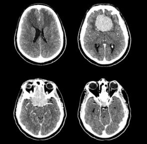
A 60-year-old right-handed male truck driver with no past medical history presented to eye clinic after six months of progressively worsening vision in the right eye, headaches, decreased sense of smell and taste, increased forgetfulness and irritability. Brain Magnetic Resonance Imaging (MRI) demonstrated a large, extra-axial mass consistent with an anterior skull base meningioma extending to the sellar and suprasellar region. Pre-op, ophthalmologic exam revealed OD hand motion detection only and OS 20/100 (Figure 1 and Figure 2). Pupils were sluggishly reactive to light bilaterally, and an OD afferent pupillary defect was present. Optic disk pallor was present in OD > OS. Slit lamp exam revealed bilateral lens nuclear sclerosis and cortical spokes. Automated perimetry showed decreased sensitivity throughout the visual field, including centrally, in OD, and temporally in OS. Visual field testing was consistent with sector scotoma bilaterally. The patient underwent an extensive 16-hour microsurgical resection with a cranio-orbito-zygomatic approach to the anterior cranial fossa and had intraoperative monitoring with somatosensory and motor evoked potentials. Bilateral A1 segments were visualized during surgery. The right Recurrent Artery of Heubner was engulfed in the tumor and was carefully dissected free and untethered from its encasement. The tumor was adherent to the optic nerves, and was invading the optic canal bilaterally and severely compressed the right greater than the left optic nerve. Simpson grade 1 resection, defined as macroscopically complete tumor resection with removal of affected dura & underlying bone, was achieved [1].

    Figure 1: Axial CT Head with contrast images demonstrating a 5.6 cm
transverse of 5.5 cm APextra-axial meningioma. Edematous changes are
seen within the frontal lobes associated with an enhancing frontal mass. The
mass occupies the anterior cranial fossa floor. The mass appears to extend
over the right-sided sphenoid ridge, might extend over the left sphenoid ridge,
and likely extends into the sella.

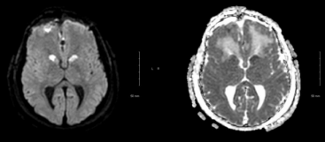
    Figure 2: Axial DWI and ADC demonstrating acute bilateral recurrent artery
of Heubner infarct. Axial DWI hyperintensity of the caudate head. B: Axial
ADC hypointensity of caudate head.
There were no immediate peri-operative complications and no intra- or peri-operative hypotension. Intra-operatively, Mean Arterial Pressure (MAP) readings were stable. Post-operatively, he remained intubated secondary to agitation and an inability to follow commands for 2 days. On day 0, patient required propofol 50 mcg/kg/min and fentanyl 100 mcg/hr. He was disoriented, violent and thrashing in bed. He required a nicardipine infusion overnight for SBP less than 140. Vital signs remained stable, and he had no signs of infection, thyroid abnormalities, or arterial blood gas abnormalities to explain his alteration in mental status. CBC and BMP were normal. A post op MRI revealed bilateral infarcts of the caudate head in the territory of the Recurrent Artery of Heubner (RAH). After extubation, patient demonstrated left hemianopsia and a right superior quadrantanopia with color desaturation. Patient was alert, attentive, interactive, and had a full affect.
At 1-month post-operative appointment, he was recovering with persistent poor vision, normal subjective sense of smell and improving cognition. Patient did not experience any abnormal movements or paresis. Three months post-operatively, he continued to have visual complaints, and was unable to return to work.
Discussion
We describe a patient who suffered bilateral RAH territory (caudate head) infarcts after resection of a large skull base olfactory groove meningioma. Our patients infarct were not related to either vasospasm or injury to perforating arteries. The surgical approach used in our case was based on the Neurosurgeons preference, tumor pathology/type, extent and its involvement of surrounding structures i.e optic nerves.
Bilateral RAH infarctions have rarely been reported. The Recurrent Artery of Heubner, commonly a branch of the proximal A2 segment (post-communicating segment) of the anterior cerebral artery, supplies the anterior limb of the internal capsule, anterior caudate and putamen, and the tip of the outer segment of the globus pallidus. Infarction typically results in contralateral paresis of the arm and face [2]. A variety of other symptoms that have been reported range from unilateral or bilateral choreiform movements, abulia, attention disorder, impaired recent memory, apathy, decreased spontaneity, depression, dementia, psychiatric self-inactivation and akinetic mutism [3-15]. These symptoms can be acute or subacute [9]. Our patient only had post-operative agitation, decreased spontaneity without other extrapyramidal complications at the three-month follow-up appointment. Approximately 21 cases of bilateral RAH infarctions have been reported [3,4,6,8,9,13]. Etiology of infarction in these cases includes variant anatomy such as a missing A1 segment [9] and vasospasm secondary to subarachnoid hemorrhage [3,15]. Olfactory groove meningiomas often extend and encase surrounding blood vessels; ACA involvement is common and can make surgical resection difficult. However, operative or perioperative ischemia remains a relatively rare complication of meningioma [16,17]. Preoperative planning with Magnetic resonance Angiogram (MRA) is not a standard of care but can be used to better identify anatomical variants of the ACAs prior to surgery.
Conclusion
Bilateral RAH infarction although rare, has been reported in the literature in association with vascular anomalies and stroke risk factors. We describe the first case of a patient who suffered bilateral infarcts of the RAH in association with a giant meningioma resection of the olfactory groove resulting in delirium requiring high doses of sedation. Cerebral infarction involving the ACA territories remains a known adverse complication of these procedures but has not been described involving the RAH bilaterally. This case report adds a new complication of olfactory meningioma resection and provides a new etiology of bilateral RAH.
References
- Sughrue ME, Kane AJ, Shangari G, et al. The relevance of Simpson Grade I and II resection in modern neurosurgical treatment of World Health Organization Grade I meningiomas. J Neurosurg. 2010; 113: 1029-10 35.
- Brazis, Masdeu JC, Biller J. Localization in Clinical Neurology. Lippincott Williams & Wilkins; 2011.
- Den heijer T, Ruitenberg A, Bakker J, Hertzberger L, Kerkhoff H. Neurological picture. Bilateral caudate nucleus infarction associated with variant in circle of Willis. J NeurolNeurosurgPsychiatr. 2007; 78: 1175.
- Goldblatt J, White NW, Wright MG. Bilateral chorea associated with caudate nuclei lacunar infarcts. A case report. S Afr Med J. 1989; 75: 443-444.
- Kumral E, Evyapan D, Balkir K. Acute caudate vascular lesions. Stroke. 1999; 30: 100-108.
- Richfield EK, Twyman R, Berent S. Neurological syndrome following bilateral damage to the head of the caudate nuclei. Ann Neurol. 1987; 22: 768-771.
- Rodier G, Tranchant C, Mohr M, Warter JM. Neurobehavioral changes following bilateral infarct in the caudate nuclei: a case report with pathological analysis. J Neurol Sci. 1994; 126: 213-218.
- Weise D, Weise G, Solymosi L, Classen J. Generalized choreic movement disorder due to bilateral recurrent artery of Heubner infarctions. Basal Ganglia. 2012; 2: 153-155.
- Fukuoka T, Osawa A, Ohe Y, Deguchi I, Maeshima S, Tanahashi N. Bilateral caudate nucleus infarction associated with a missing A1 segment. J Stroke Cerebrovasc Dis. 2012; 21: 908.e11-12.
- Kuriyama N, Yamamoto Y, Akiguchi I, Oiwa K, Nakajima K. Bilateral caudate head infarcts. RinshoShinkeigaku. 1997; 37: 1014-1020.
- Mendez MF, Adams NL, Lewandowski KS. Neurobehavioral changes associated with caudate lesions. Neurology. 1989; 39: 349-354.
- Mrabet A, Mrad-ben hammouda I, Abroug Z, Smiri W, Haddad A. Bilateral infarction of the caudate nuclei. Rev Neurol Paris. 1994; 150: 67-69.
- Lim JK, Yap KB. Bilateral caudate infarct--a case report. Ann Acad Med Singap. 1999; 28: 569-571.
- Trillet M, Croisile B, Tourniaire D, Schott B. Disorders of voluntary motor activity and lesions of caudate nuclei. Rev Neurol Paris. 1990; 146: 338-344.
- Mizuta H, Motomura N. Memory dysfunction in caudate infarction caused by Heubner's recurring artery occlusion. Brain Cogn. 2006; 61: 133-138.
- Pallini R, Fernandez E, Lauretti L, et al. Olfactory groove meningioma: report of 99 cases surgically treated at the Catholic University School of Medicine, Rome. World Neurosurg. 2015; 83: 219-231.e1-3.
- Zygourakis CC, Sughrue ME, Benet A, Parsa AT, Berger MS, Mcdermott MW. Management of planum/olfactory meningiomas: predicting symptoms and postoperative complications. World Neurosurg. 2014; 82: 1216-1223.