Case Report
Austin J Clin Neurol 2017; 4(1): 1099.
Tolosa-Hunt Syndrome with Involving Structures outside the Cavernous Sinus: Report a Case with Histological Study
Belfquih H* and El Mostarchid B
Department of Neurosurgery, Mohammed V Military Teaching Hospital, Morocco
*Corresponding author: Hatim Belfquih, Department of Neurosurgery, Mohammed V, Military Teaching Hospital, Rabat, Morocco
Received: November 10, 2016; Accepted: January 24, 2017; Published: January 27, 2017
Abstract
Subject: To report a case of Tolosa-Hunt syndrome (THS) with involving structures outside the cavernous sinus (CS) with histological confirmation.
Case Presentation: A 37 year immuno-competent man presented with right unilateral painful ophthalmoplegia. Computer Tomodensitometry scan (CT) and Magnetic resonance imaging (MRI) showed a lesion of right CS with extension in to upper clivus and posterior wall of CS. Exhaustive biological and radiological study showed no abnormalities and excluded others aetiologies mimicking THS. The involving outside of CS was uncommon. A surgical biopsy was made via transsphenoidal approach targeting the posterior extension of the SC lesion in the upper clivus. Histological study showed an idiopathic non granulomatous inflammation. The response to systemic corticosteroid therapy was positive with clinical and radiological follow up.
Conclusion: The aetiology of THS is an idiopathic non granulomatous inflammation process of CS. The diagnosis is therefore made by exclusion. Biopsy of the lesion may require confirming the diagnosis. Biopsy of the posterior wall of cavernous sinus via transsphenoidal approach is simple rapid and effective to confirm the non-specific inflammatory in THS and excluding others aetiologies mimicking THS.
Keywords: Tolosa-Hunt syndrome; Magnetic resonance imaging; Biopsy; Transsphenoidal approach; Idiopathic non granulomatous inflammation
Introduction
Tolosa-Hunt syndrome (THS) also known as painful ophthalmoplegia is a rare entity characterised by unilateral retroorbital or hemicranial pain and variable numbness and paresis of the third, fourth, and or sixth cranial nerves. Having a relapsing and remitting course and response to systemic corticosteroid therapy [1,2].
THS is due to idiopathic non granulomatous inflammation process of Cavernous Sinus (CS). The diagnosis is made by exclusion of others infiltrative processes and infection processes that mimic THS [3,4].
A case of THS involving structures outside the cavernous sinus with histological confirmed is reported.
Case Presentation
28 year old male was admitted for punt full ophthalmoplegia. Four weeks prior to admission, the patient developed right retroorbital and forehead pain. He denied any recent trauma or HIV risk factors. Two weeks prior to admission, he developed binocular diplopia in all directions of gaze, associated with a feeling of dizziness with neuralgia in right V1 and proptosis.
Motor and sensory examination was normal. Ophthalmologic evaluation showed: That visual acuity was 9/10 in right and left eye. Pupils are 3mm and reactive to light. A right ophthalmoplegia was noted with ptosis and weakness in the right. Segment anterior and posterior were normal. Fundi and visual field were normal. An hypoesthesia in right V1, V2 distribution with decreased cornien reflex. Smile is symmetrical, hearing intact. Coordination evaluation was unremarkable. Oto-rhyno-laryngologic evaluation was normal. The somatic examination was unremarkable. Electrolytes and glucose were normal. CBC count showed mild leukocytosis to 15.1 00 / m3. Erythrocyte sedimentation rate was 40 first hour. CR protein was 63 mg/L. Fibrinogen was 5, 19 g/l. Haptoglobine was 2, 77 g/l (0-3,2 g/l). Orosumucoïde 1,1 g/l (0,5 – 1,3 g/l). Lyme titre was negative (anti-corps anti-B Burgdorferi Ig G (reactif EIA Behring°). Fluorescent treponema antibody, TPHA and VDRL were negatives. HIV is negative. Lupus erythematous preparation was negative. Hepatic evaluation was normal, Antinuclear antibody was negative. Intradermo-reaction for 10% tuberculin was 6 mm to 72 hours control. Cerebrospinal fluid study by lumbar punction was normal.
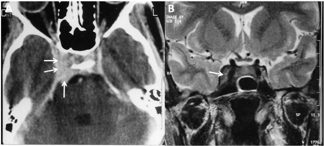
Cranial Computer Tomography (CT) scan revealed increased density in the right cavernous sinus and upper clivus (Figure 1A). Magnetic Resonance Imaging (MRI) showed cavernous sinus lesion extending to the Gasserian ganglion from the CS and spreading into clivus (Figure 1B). Angio-MRI showed no vascular lesions or intracavernous carotid anomalies in the cavernous sinus. The diagnosis of TH syndrome was done but extra CS extension was not common.

    Figure 1: CT scan of brain on axial (A) after intravenous infusion of contrast
showing enlarged right cavernous sinus (arrows). Coronal T2-weighted MRI
image (B) demonstrates non specific fullness involving the right cavernous
sinus consistent with the THS within the context of the history (arrow).
Since his ophthalmoplegia had progressed rapidly and excluding others aetiologies mimicking THS, A surgical biopsy was performed via translabial transsphenoidal approach to the clivus and posterior wall of right cavernous sinus. Exploration revealed a thin layer of grayfish red hemorrhagic granulation tissue covering clivus and the posterior right wall of CS. Histologic examination showed non granulomatous chronic inflammatory tissue containing polymorphonuclear cells. Bacteriological study showed no germ and no grows. The THS was suspected and the patient was treated with intravenous Methyl Prednisolone: 120 mg/day during ten days following by a 60mg dose of oral prednisone during two months. Within 72 hours the patient responded with resolution of his retroorbital pain and gradual improvement in ptosis and ophthalmoplegia.
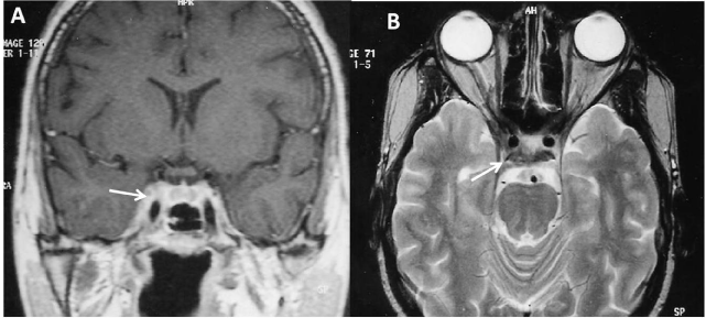
Fourth month postoperative MRI showed near normal size of right cavernous sinus (Figure 2). The patient is asymptomatic with 36 months follow-up.

    Figure 2: Fourth month Coronal T1-weighted MRI image (A) and axial (B)
after intravenous infusion of contrast demonstrates near normal size of right
cavernous sinus.
Discussion
THS is a complex of periorbital or hemicranial pain associated with ipsilateral ophthalmoplegia, oculosympathetic dysfunction, and sensory loss in the first division of the trigeminal nerve. The aetiology is an idiopathic non granulomatous inflammation process. The diagnosis is therefore, and by exclusion [1,2].
In this case the diagnosis of the THS was made by exclusion of others aetiologies and histological confirmation of non granulomatous inflammation process. Such observations with histological study are rarely reported in the literature.
The original criteria of THS were reported in 1966. In 1988, Hunt W. E and. Brightman R.P [3] proposed a revision of the original criteria to include cases meeting all other criteria for THS but showing involvement of V2, V3 or Cranial nerve VII (witch lie outside of the sinus cavernous sinus). Their criteria therefore included cases « rarely involving structures outside the CS ». In addition, these authors downplayed the need for surgical exploration.
Contrast enhanced MRI with multiple views, should be the initial diagnostic study performed. Numerous reports have demonstrated an area of abnormal soft tissue in the region of the CS in most, but not all, patients with THS. The typically, the abnormality is seen an intermediate signal intensity on T1 and intermediate weighted images, consistent with an inflammatory process. In addition, there is enhancement of the abnormal area after intravenous injection of paramagnetic contrast [1,5].
Cerebral angiography can detect abnormalities in the intracavernous carotid artery in patient with THS. These have been described as « segmental narrowing » or constriction [1,2]. These changes are not specific to THS. Because of this fundamental limitation of initial imaging studies, some authorities would suggest that resolution of imaging abnormalities after a course of systemic corticosteroids should be considered of THS. Ohta S and all [6] had reported a bilateral cavernous sinus actinomycosis resulting in painful ophthalmoplegia.
Neurosurgical biopsy is rarely employed to establish the diagnosis. This can be technically difficult. It usually involves biopsy of the dural wall of SC. We have target the posterior extension of the SC lesion in the upper clivus via transsphenoidal approach. This procedure was more simple rapid and effective to confirm the nonspecific inflammatory in our patient.
References
- Areaya AA, Cerezal L, Canga A, Polo JM, Berciano J, Pascual J. Neuroimaging diagnosis of Tolosa-Hunt syndrome: MRI contribution. Headache. 1999; 39: 321-325.
- Forderreuther S, Straube A. The criteria of the International Headache Society for Tolosa-Hunt syndrome need to be revised. J Neurol. 1999; 246: 371-377.
- Hunt WE, Brightman RP. The Tolosa-Hunt syndrome: A problem in differential diagnosis. Acta Neurochir Suppl(Wien) 1988; 42: 248-252.
- Kline LB, WF Hoyt. The Tolosa-Hunt syndrome. J Neurol Neurosug Psychiatry. 2001; 71: 577-582.
- Vallat JM, Vallat M, Julien J. Painful ophtalmoplegia (Tolosa-Hunt) accompanied by peripheral facial paralysis. Ann Neurol. 1980; 8: 645.
- Ohta S, Nishizawa S, Namba H, Sugimura H. Bilateral cavernous sinus actinomycosis resulting in painful ophtalmoplegia. Case report. J Neurosurg. 2002; 96: 600-602.