Case Report
Austin J Clin Neurol 2017; 4(2): 1103.
A 47-Year-Old Woman with Seizure and Rapidly Progressive Dementia
Haeri G, Razmeh S, Almasi-Dooghaee M*, Habibi AH, Sina F and Ghurchian Z
Department of Neurology, Iran University of Medical Sciences, Iran
*Corresponding author: Mostafa Almasi-Dooghaee, Department of Neurology, Iran University of Medical Sciences, Rasoul-e-Akram Hospital, Niayesh St, Sattarkhan ST, Tehran, Iran
Received: January 24, 2017; Accepted: March 16, 2017; Published: March 24, 2017
Abstract
Paraneoplastic encephalitis is a rare autoimmune inflammatory disorder which can be associated with different malignancies. Its common symptoms are personality and behavioral disorders, seizure, confusion, mental decline and abnormal movements. It may manifest itself before the presentation of underlying malignancy and can cause permanent neurologic sequels despite treatment of cancer. It is recommended that the physician should be aware of the ambiguous symptoms of paraneoplastic disorders to perform investigations in order to find the true diagnosis and treat promptly. We report a patient with seizure and rapidly progressive dementia with a diagnosis of anti-voltage-gated potassium channel (VGKC) encephalitis as the first presentation of small cell lung carcinoma (SCLC).
Keywords: Small cell lung carcinoma; Paraneoplastic encephalitis; Anti voltage-gated potassium channel
Introduction
Paraneoplastic encephalitis is an autoimmune inflammatory disorder of the central nervous system and is associated with different malignancies including breast, testis, bladder and thymus tumors and lymphoma; however, approximately eighty percent of reported cases are due to small cell lung cancer (SCLC) [1,2]. It usually occurs at middle age with a subacute onset and steady progression over a period of weeks to months. The usual symptoms and signs are sleep and personality disorders, depression, anxiety, partial and generalized seizures, hallucination, delusion, cognitive decline and abnormal movements [3]. This syndrome may precede the diagnosis of malignancy by months or years; In other words, it can present rarely as the first manifestation of cancer [4]. We present a case of paraneoplastic limbic encephalitis with antibody against voltagegated potassium channel as a first manifestation of SCLC that was followed for several months.
Case Presentation
A 47- year-old Iranian woman was admitted in neurology ward of Rasoul Akram hospital due to frequent generalized tonic seizures. The patient developed fever, headache, agitation, paranoid hallucination and frequent seizures about 40 days prior to admission in our hospital and was treated with the diagnosis of herpes encephalitis in another medical center but the lack of improvement led to referring to our hospital for further investigations. She didn’t take any medications before beginning of symptoms and her family history and review of systems were noncontributory. She denied any history of seizures before this presentation.
On physical examination, she was awake and the vital signs were normal. She had normal heart rate and regular cardiac rhythm and no cardiac murmur was detected. The lungs had not any significant findings on examination. The breasts examination was normal without any rash, mass, or other abnormal signs. The abdomen was soft with normal size of liver and spleen. Also, the Extremities had not any remarkable findings.
In the neurological examination, the Mini-Mental State Examination (MMSE) score was 20 out of 30. In details, the sub scores of registration, attention, calculation and recall were impaired. All of the cranial nerves were completely normal. Muscle strength and all deep tendon reflexes were normal and plantar responses were flexor bilaterally.
Fluid-attenuated inversion recovery (FLAIR) and T2-weighted MRI scan of brain revealed high intensity changes in bilateral medial temporal lobes (Figure 1). The routine cerebrospinal fluid (CSF) parameters including leukocyte count and protein were in normal range and the result of polymerase chain reaction (PCR) for herpes simplex DNA in CSF was negative, on two separate occasions. Electroencephalogram (EEG) showed diffuse background slowing and scattered epileptiform discharges including generalized spike and wave complexes.

    Figure 1:  Axial FLAIR (A) and T2 (B) sequences of brain MRI showed hyperintensity signals in bilateral medial temporal lobes without volume loss or mas effect.
Diffusion-weighted and apparent diffusion coefficient (ADC) sequences showed hyper signal changes (C and D).
The paraneoplastic encephalitis was suspected due to the clinical features and paraclinical findings [5,6]. The investigations for paraneoplastic disorders were conducted including chest and abdominopelvic CT scans and mammography that all were normal. The paraneoplastic antibodies had been assessed in the blood serum including antibodies against NMDA receptor, VGKC, Yo, Ri, Hu, Ma1, Ma2, and amphiphysin. Among these tests, just the anti-VGKC antibody was positive.
The patient treated with 0.4 gr/kg/day for five consecutive days (total dose: 120 gr) intravenous immuno globulin (IVIG) with the diagnosis of autoimmune anti-VGKC encephalitis. The seizures were ceased and the cognitive functions including behavioral changes were clearly improved clinically. Finally the patient discharged with prescription of valproate sodium 500 mg twice daily.
One month later, the patient admitted again with status epilepticus and treated with intravenous phenytoin and valproate sodium and oral levetiracetam. Also, she developed hyponatremia during the course of admission. The diagnosis of syndrome of inappropriate antidiuretic hormone secretion (SIADH) was suggested by nephrology consult after excluding the other causes of hyponatremia. After the correction of serum sodium levels and control of seizures, the patient was discharged. Regular follow-up after discharge showed that the seizures were completely controlled.
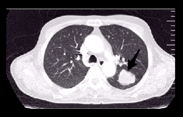
Six months after discharge, she developed a productive persistent cough. The investigations including chest CT scan revealed a hyperdense lesion in the left lung (Figure 2). Finally, the biopsy showed SCLC and the patient was referred to chemotherapy unit.

Figure 2: Axial chest CT scan revealed a hyperdense mass in the left lung.
Discussion
The first report of a patient with psychiatric disorders associated with small cell lung carcinoma was described by Corelli’s at 1956 [7]. He invented a term for this disorder and called it “limbic encephalitis” (LE) [8]. Afterward, different kinds of malignancies were reported with this syndrome; however the major cause of this syndrome remained lung cancer especially SCLC. Lawn and his colleagues reviewed 24 patients suspicious to limbic encephalitis that 13 patients had small cell lung carcinoma [9]. Their study showed that the common presentations of limbic encephalitis were intellectual dysfunctions, seizures, and psychiatric disorders. Antineuronal antibodies were positive in 14 out of 22 patients and all of the patients had the abnormal electroencephalogram (EEG) [9]. The reported patient in this study had no abnormality in routine CSF parameters but in Lawn and coworkers’ study, the CSF abnormalities were detected in 18 of 23 patients [9]. Their findings showed that pleocytosis, elevated protein and presence of oligoclonal band were detected in the CSF analysis of majority of the patients.
The most important investigations for diagnosis of limbic encephalitis were belonged to autoimmune antibodies. Anti-Hu is the most common antibody and is detected in approximately half of the paraneoplastic encephalitis [10]. It was negative in our patient. In a study, that done by Alamo witch and colleagues retrospectively, it was shown that the underlying malignancy should be considered even if the anti-Hu antibodies were negative in patients with suspicious to LE [11]. They also suggested that the patients with LE and SCLC who had not the anti-Hu antibody, are more responsive to treatment of cancer [11].
Another antibody is anti-VGKC antibody that was positive in our case. Its manifestations in nervous system are very diverse including limbic encephalitis with seizure, dementia, hallucination and psychiatric symptoms, sleep disorders (hypersomnia, insomnia, REM sleep behavior disorder), peripheral nerve hyperexcitability and autonomic dysfunction (Morvan’s syndrome) [12,13]. One of the other unusual manifestation of LE is hyponatremia which was present in this reported patient. In a study, Adedayo and colleagues reviewed the different causes of patients with hyponatremia and demonstrated malignancies especially SCLC as one of the important causes of hyponatremia [14]. They suggested that early detection and treatment of this fatal condition, can improve the patient’s prognosis [14].
In Conclusion, paraneoplastic encephalitis is rare disorder associated with different malignancies especially SCLC that can be present with personality and behavioral changes, seizure and cognitive decline. It may develop several months before the diagnosis of malignancy and may be misdiagnosed with other conditions if it is not considered.
References
- Blaes F. Paraneoplastic neurological syndromes--diagnosis and management. Curr Pharm Des. 2012; 18: 4518-4525.
- Shirafuji T, Kanda F, Sekiguchi K, Higuchi M, Yokozaki H, Tanaka K, et al. Anti-Hu-associated paraneoplastic encephalomyelitis with esophageal small cell carcinoma. Intern Med. 2012; 51: 2423-2427.
- Adam VN, Marin D, BudinceviÄ H, MrsiÄ V, GoranoviÄ T, TonkoviÄ D. Paraneoplastic limbic encephalitis. Acta Med Croatica. 2012; 66: 29-32.
- Tüzün E, Dalmau J. Limbic encephalitis and variants: classification, diagnosis and treatment. Neurologist. 2007; 13: 261-271.
- Graus F, Saiz A, Lai M, Bruna J, López F, Sabater L, et al. Neuronal surface antigen antibodies in limbic encephalitis: clinical-immunologic associations. Neurology. 2008; 71: 930-936.
- Foster AR, Caplan JP. Paraneoplastic limbic encephalitis. Psychosomatics. 2009; 50: 108-113.
- Charatan FB, Brierley JB. Mental disorder associated with primary lung carcinoma. Br Med J. 1956; 1: 765-768.
- Corsellis JA, Goldberg GJ, Norton AR. “Limbic encephalitis” and its association with carcinoma. Brain. 1968; 91: 481-496.
- Lawn ND, Westmoreland BF, Kiely MJ, Lennon VA, Vernino S. Clinical, magnetic resonance imaging, and electroencephalographic findings in paraneoplastic limbic encephalitis. Mayo Clin Proc. 2003; 78: 1363-1368.
- Sabin TD, Jednacz JA, Staats PN. Case records of the Massachusetts General Hospital. Case 26-2008. A 26-year-old woman with headache and behavioral changes. N Engl J Med. 2008; 359: 842-853.
- Alamowitch S, Graus F, Uchuya M, Reñé R, Bescansa E, Delattre JY. Limbic encephalitis and small cell lung cancer. Clinical and immunological features. Brain. 1997; 120: 923-928.
- Vincent A, Buckley C, Schott JM, Baker I, Dewar BK, Detert N, et al. Potassium channel antibody-associated encephalopathy: a potentially immunotherapyresponsive form of limbic encephalitis. Brain. 2004; 127: 701-712.
- Irani SR, Michell AW, Lang B, Pettingill P, Waters P, Johnson MR, et al. Faciobrachial dystonic seizures precede Lgi1 antibody limbic encephalitis. Ann Neurol. 2011. 69: 892-900.
- Onitilo AA, Kio E, Suhail AR. Tumor related hyponatremia. Clin Med Res. 2007; 5: 228-237.