
Case Presentation
Austin J Clin Neurol 2021; 8(1): 1143.
The Etiology and Secondary Prevention of a Stroke Patient after Successful Catheter Ablation
Zhao J*
Department of Neurology, China
*Corresponding author: Jing Zhao, Department of Neurology, Xinsong Road 170, Shanghai, China
Received: January 25, 2021; Accepted: February 22, 2021; Published: March 01, 2021
Abstract
Ischemic Stroke (IS) is one of the leading diseases of mortality and disability worldwide. The rational administration of anticoagulant or antiplatelet drugs is of great importance to prevent stroke recurrence. Here we report an IS patient with a medical history of Catheter Ablation (CA) of Atrial Fibrillation (AF), who had a single vessel scattered lesions in the head Magnetic Resonance Imaging (MRI) scan, which indicated a remarkable correlation with morphology of atherosclerotic artery and artery embolism, while no AF recurrence was detected. At last, we focused on the possible criminal artery and found vulnerable plaques from apparent positive remodeling demonstrated by High-Resolution Vessel Wall MRI (HRVW-MRI). We provided a secondary prevention with the statin and antiplatelet therapy to reduce the risk of bleeding caused by anticoagulation and asked him to follow up Holter every three months. Along with this case report, we describe the imageology, review of literature and treatment outcomes in regard to CA of AF and discuss the prevention of IS.
Keywords: Ischemic stroke; Catheter ablation; Atrial fibrillation; HRVW-MRI
Case Presentation
A 72-year-old man woke up with left side numbness and weakness, slight alalia and the angle of his mouth deviated to right side. The patient arrived at our emergency department one hour later, as his left limb numbness disappeared, muscle strength and speech returned to normal. The neurologic examination demonstrated that left nasolabial groove was slightly shallower and left Babinski sign was positive, while the muscle strength and muscular tension of limbs were normal. Computed Tomography (CT) showed the residual left temporooccipital encephalomalacia. The National Institutes of Health Stroke Scale (NIHSS) score was 1. The patient reported a history of hypertension and atherosclerosis, with blood pressure controlled around 140-150/80-90mmHg. He also had a history of cerebral infarction 4 years ago when AF was found. CA of AF was performed 2 years ago and oral administration of aspirin was given after surgery. The patient didn’t feel palpitation and the every threemonth checking Holter showed no AF rhythm. A few weeks before this onset, the patient took bisoprolol by himself, and his lowest heart rate was 40bpm with occasional dizziness. The admission diagnosis is cerebral infarction (OCSP: PACI); grade hypertension; personal history of CA of AF (CHA2DS2-VASc score of 4). Aspirin and statins was given while thrombolytic therapy wasn’t performed due to the low NHISS score and awaken stroke.
After admission, laboratory tests (fasting plasma glucose, plasma glucose of two hours after meal, glycated hemoglobin, blood lipids, homocysteine, autoimmune antibodies, etc.) were almost normal. Holter showed some atrial premature beats, and no AF rhythm was caught. Echocardiography (EEG) showed a slightly enlarged left atrium (41mm) and a slightly thickened interventricular septum (12mm). The head Magnetic Resonance Imaging (MRI) showed some scattered diffuse limited lesions in the cortical and subcortical areas around the right central sulcus (Figure 1A-1D). The head and neck Computed Tomography Angiography (CTA) showed a suspicious stenosis in M2 segment of right Middle Cerebral Artery (MCA), which was the criminal artery comprised of precentral artery, artery of central sulcus and artery of postcentral sulcus, as well as a moderate non-criminal stenosis in M1 segment of left MCA (Figure 1E).
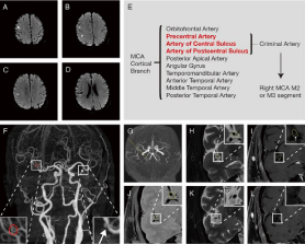
    Figure 1: (A-D) MRI showed the diffuse limited lesions (the white lesion) are
scattered in the cortical and subcortical areas near the right central sulcus.
(E) Diagram of the criminal artery. (F) Head and neck CTA of patient showed
a suspicious criminal stenosis in M2 segment of right MCA (the red circle
shows), and a moderate non-criminal stenosis in M1 segment of left MCA (the
arrow shows). (G) The orientation MRA of figure H, I, J, K and L. (the yellow
line) (H) The T2WI sequence of HRVW-MRI shows artery outline (the circle
shows) and the plaque mixed single (the circle shows). (I) The enhanced
TIWI sequence of HRVW-MRI shows the outline (the circle shows) and the
plaque slightly enhanced single (the arrow shows). (J) The PDWI sequence
of HRVW-MRI shows artery outline (the circle shows) and the plaque mixed
single (the arrow shows). (K) The T2WI sequence of HRVW-MRI of other
lay shows no artery plaque mixed single around the artery outline. (L) The
enhanced TIWI sequence of HRVW-MRI of other layer shows no artery
plaque mixed single around the artery outline. Due to the slight movement
of the patient during examination, the layers of H, I, J and K don’t overlap
completely.
Given the diffuse limited lesions in head MRI, a suspicious criminal artery stenosis and no AF recurrence evidence, the etiology of stroke might be atherosclerotic embolism. To find more evidence to differentiate cardiogenic embolism and artery-to-artery embolism, a High-Resolution Vessel Wall MRI (HRVW-MRI) was implemented and consequently a positive remodeling was found (Figure 1C). The Chinese Ischemic Stroke Subclassification (CISS) typing was Atherosclerotic (artery-to-artery embolization + low emboli clearance rate).
Diffusion-Weighted Imaging (DWI) sequence plays an important role in the diagnosis of cerebral infarction with high positive predictive value to confirm diagnosis and indicate the etiology and classification of stroke. The study of lesion pattern and cerebral infarction etiology showed that multiple lesions are more common in Large- Artery Atherosclerosis (LAA), while large area cerebral infarction, or the lesions involving the anterior and posterior circulation at the same time is more common in cardiac etiology [1]. The single vessel scattered infarcts are more correlated with atherosclerotic vascular disease [2]. Therefore, the etiology of the stroke might be atherosclerotic embolism rather than cardiac embolism.
By HRVW-MRI, we took comprehensive consideration and identified the cause of IS as atherosclerosis of vessel wall. No fluctuation conditions was observed during hospitalization. The secondary prevention of cerebral infarction consisted of antiplatelet agents, statins, blood pressure control, diet control and improvement of lifeway. Unexpectedly, one month after this stroke onset, the patient developed palpitation and presented to the emergency department of our hospital, with his ECG indicating atrial flutter (2:1 conduction) and paroxysmal supraventicuar tachycardia. The patient received CA again and maintained long-term oral anticoagulation with dabigatran and statin. No recurrence of atrial fibrillation and cerebral infarction was seen during regular follow-up.
Discussion
In this case, an IS patient with a medical history of CA of AF had a single vessel scattered lesions in MRI scan, which indicated a remarkable correlation with morphology of atherosclerotic artery and artery embolism. Meanwhile, no evidence of AF recurrence was obtained. At last, we focused on the possible criminal artery and found vulnerable plaques through apparent positive remodeling by HRVWMRI. We further suggested the patient to accept transesophageal echocardiography in order to exclude mural thrombus, but the patient rejected. However, in this case, considering atherosclerosis as etiology, we provided a secondary prevention with the statin and antiplatelet therapy to reduce the risk of bleeding caused by anticoagulation and asked him to follow up Holter every three months. Although the patient received CA again and maintained long-term anticoagulation due to the arrhythmia presented with palpitation afterwards, an inexistent symptom in the course of this stroke, we regarded atherosclerosis as the cause of the stroke, because the unstable plaque could fully explain the lesion and no symptom or evidence of arrhythmia was detected during this onset to establish the causal relationship between arrhythmia and this onset. The atherosclerosis might be a possible explanation of the results obtained by ESUS trial that anticoagulation was not superior to antiplatelet for secondary prevention after embolic stroke from undetermined source, as undetected paroxysmal atrial fibrillation was not a major cause of recurrent stroke [3,4].
The etiological classification plays an essential role in the diagnosis and treatment of acute cerebral infarction, which implies the possibility of more individual and reasonable prevention protocols. Head MRI has been widely used in clinic, which accurately shows the location, size and distribution of diffused restricted lesions, and gives a clue for the etiological classification [1,2]. Combined with laboratory and imaging examinations, a more rational stroke classification and a more reasonable secondary prevention scheme can be obtained. The common etiology, such as atherosclerosis, oval foramen defect and AF, may exist at the same time [5]. With the development of imaging technology, HRVW-MRI provides us with more information about vascular wall and plaque [6]. Therefore, we may give patients a more appropriate prevention plan.
Vascular remodeling is the changes in vascular wall structure to adapt to hemodynamic changes, including positive remodeling and negative remodeling [7].
Positive remodeling
• The diameter of vessels increases significantly, and the lumen area does not shrink or slightly increases.
• It mostly occurs at the early stage of atherosclerotic lesion, which contains more inflammatory factors and new blood vessels. It is easy to associate with hemorrhage in the plaque and contributes to the formation of vulnerable plaque.
• There is no significant stenosis in the lumen, which cannot be found by CTA, Magnetic Resonance Angiography (MRA), Digital Subtraction Angiography (DSA) or other lumen examinations when positive remodeling happens.
Negative remodeling
• When negative remodeling happens, there is no significant change or increase in vessel diameter, but the lumen area decreases typically.
• The area of the cavity is obviously reduced, and more stable than that in the positive remodeling.
In 2019, Wang et al. found that HRVW-MRI could identify the undetectable high-risk plaque features and detect the abnormity of the vascular wall, such as wall enhancement, positive remodeling, intraplaque hemorrhage, plaque location and eccentricity [8]. Thus, HRVW-MRI is a promising tool to stratify IS patients, which is of great significance for more reasonable etiological classification and secondary prevention.
It is controversial whether anticoagulation is necessary for patients after successful CA. A meta-analysis showed that no statistical difference was seen in the incidence of thromboembolism between long-term anticoagulation and non-anticoagulation patients after CA of AF (OR 1.83, 95% CI 0.69-4.88; p=0.221), while the risk of bleeding was significantly decreased in the non-anticoagulation group (OR 6.5, 95% CI 1.93-21.86; p=0.002), which raised doubts about the net clinical benefit of long-term anticoagulation [9]. Given the increased risk of bleeding and death caused by anticoagulation therapy, we suppose that regular follow-up of Holter and the conventional secondary prevention of antiplatelet therapy is reasonable for IS patients with vulnerables plaque in the criminal artery and without the recurrent AF after successful CA. The ongoing trial, Optimal Anti-Coagulation for Enhanced-Risk Patients Post- Catheter Ablation for Atrial Fibrillation (OCEAN), is expected to be completed by December 2021, the results of which may guide future antithrombotic strategies [10].
Although the patient takes long-term dabigatran in the end, we think it may be reasonable to select antiplatelet drugs as the secondary prevention for IS patients after successful CA of AF, when the evidence of unstable atherosclerosis in the criminal artery is found rather than that of AF recurrence or mural thrombus. Detailed pathophysiological information of patients can be obtained through various examinations to clarify the etiology of cerebral infarction and guide further treatment, one of which is HRVW-MRI that provides vascular information.
References
- Roh JK, Kang DW, Lee SH, Yoon BW and Chang KH. Significance of acute multiple brain infarction on diffusion-weighted imaging. Stroke. 2000; 31: 688- 694.
- Kang DW, Chalela JA, Ezzeddine MA and Warach S. Association of ischemic lesion patterns on early diffusion-weighted imaging with TOAST stroke subtypes. Arch Neurol. 2003; 60: 1730-1734.
- Hart RG, Sharma M, Mundl H, Kasner SE, Bangdiwala SI and Berkowitz SD, et al. Rivaroxaban for Stroke Prevention after Embolic Stroke of Undetermined Source. N Engl J Med. 2018; 378: 2191-2201.
- Diener HC, Sacco RL, Easton JD, Granger CB, Bernstein RA and Uchiyama S, et al. Dabigatran for Prevention of Stroke after Embolic Stroke of Undetermined Source. N Engl J Med. 2019; 380: 1906-1917.
- Schnabel RB, Yin X, Gona P, Larson MG, Beiser AS and McManus DD, et al. 50 year trends in atrial fibrillation prevalence, incidence, risk factors, and mortality in the Framingham Heart Study: a cohort study. Lancet. 2015; 386: 154-162.
- Bodle JD, Feldmann E, Swartz RH, Rumboldt Z, Brown T and Turan TN. High-resolution magnetic resonance imaging: an emerging tool for evaluating intracranial arterial disease. Stroke. 2013; 44: 287-292.
- August P and Suthanthiran M. Transforming growth factor beta signaling, vascular remodeling, and hypertension. N Engl J Med. 2006; 354: 2721-2723.
- Wang Y, Liu X, Wu X, Degnan AJ, Malhotra A and Zhu C. Culprit intracranial plaque without substantial stenosis in acute ischemic stroke on vessel wall MRI: A systematic review. Atherosclerosis. 2019; 287: 112-121.
- Santarpia G, De Rosa S, Sabatino J, Curcio A and Indolfi C. Should We Maintain Anticoagulation after Successful Radiofrequency Catheter Ablation of Atrial Fibrillation? The Need for a Randomized Study. Front Cardiovasc Med. 2017; 4: 85.
- Verma A, Ha ACT, Kirchhof P, Hindricks G, Healey JS and Hill MD, et al. The Optimal Anti-Coagulation for Enhanced-Risk Patients Post-Catheter Ablation for Atrial Fibrillation (OCEAN) trial. Am Heart J. 2018; 197: 124-132.