Case Report
Austin J Clin Ophthalmol. 2014;1(6): 1029.
Multidisciplinary Rehabilitation for a Patient with Pituitary Adenoma and Stroke
Kar Ho Siong1, George C Woo1, Dora Yuk Lin Chan2, Hobby Cheung3, Claudia Kam Yuk Lai4 and Allen Ming Yan Cheong1*
1School of Optometry, Hong Kong Polytechnic University, China
2Department of Occupational Therapy, Kowloon Hospital, China
3Department of Rehabilitation, Kowloon Hospital, China
4School of Nursing, Hong Kong Polytechnic University, China
*Corresponding author: Allen Ming Yan Cheong, School of Optometry, The Hong Kong Polytechnic University, Hung Hom, Hong Kong, china
Received: July 10, 2014; Accepted: Aug 05, 2014; Published: Aug 06, 2014
Abstract
Purpose: To demonstrate the clinical benefits of a multidisciplinary rehabilitation program provided by Optometric rehabilitation and occupational therapy.
Case Report: A 54-year-old male patient with multiple disabilities due to primary pituitary adenoma and secondary stroke was referred for rehabilitation by occupational therapists and optometrists, where improvements were shown in physical, cognitive, and functional performances (eye-hand coordination and mobility). Improvement in the physical performance was also shown in the field expansion by wearing Fresnel prism during orientation and mobility. Enhancements in self-perceived functional performance and fewer mobility difficulties were also reported.
Conclusion: Early recognition and intervention (e.g. vision rehabilitation) of visual problems is essential for multidisciplinary management on patients with acquired brain injury for improving their quality of life.
Keywords: Stroke; Rehabilitation; Vision; Multidisciplinary Communication; Tumor
Introduction
Pituitary adenoma is noncancerous benign tumor of the pituitary gland. It is a slow-growing tumor, affecting about 16.7 % of people in the general population [1]. Patients may report various clinical symptoms such as headaches; blur vision, and changes in mood and behavior [2]. A pituitary adenoma may compress the optic chiasm and retinal nerve fibers (RNF), leading to a corresponding bitemporal visual field (VF) defect and functional disability. Prescription of prism can increase the peripheral awareness to improve independence and confidence during mobility [3]. Although the tumor can be managed by radiotherapy and surgical removal, the effect on the VF defect may not be recoverable [4]. Furthermore, the surgical management increases the potential risk of vascular complications, patient’s body coordination, physical and cognitive functions [5]. To manage patients with multiple disabilities in both visual and physical caring, rehabilitation should cover two aspects: muscle and cognition training by occupational therapists and vision rehabilitation by optometrists. In this case report, a multidisciplinary rehabilitation program was provided for a patient with multiple deficits due to pituitary adenoma and secondary stroke. The study followed the tenets of Declaration of Helsinki and informed consent was obtained from the patient.
Case Presentation
Medical history
Patient CTS (54 year-old Hong Kong Chinese male) complained of blurry vision, speech disturbance and limb weakness on the left side since December 2009. He was then referred to a neurologist for Magnetic Resonance Imaging (MRI) examination. A 3 cm X 2.8 cm X 2.5 cm macro-adenoma was diagnosed at his pituitary gland. Surgical removal of the tumor was successful. Unfortunately, secondary stroke (infarction in left lacunars region) occurred after surgery. Patient reported poor vision and he was diagnosed with temporal VF defect on right and left eye respectively after the surgery. However, no vision rehabilitation was arranged at this stage. Right hemi paresis, multiple cognitive and physical impairments were also identified and this might delay the referral to optometric rehabilitation.
Occupational rehabilitation
The patient received occupational rehabilitation from January 2010 for 6 months (two times per week), with training targeted on upper limb strength, gait performance and cognitive function. Training on trunk rotation [6], visual scanning [7], individualized activities of daily living (ADL) and instrumental activities of daily living (IADL) were also given. To evaluate the effectiveness of training, physical and cognitive functions, and functional measures on daily tasks were examined at 3 time-points: baseline (before the intervention) and post-intervention (2-month and 6-month, Table 1).
    Visit 1 (Baseline)
    Visit 2
      (2-month)
    Visit 3
      (6-month)


    Physical function


    Power grip (kg)* a
    10
    17
    19


    Nine-Hole Peg test    (sec)* b
    85
    66
    50


    Strength of lateral    pinch a
    6
    8
    10


    Functional Test for    the Hemiplegic Upper Extremity (FTHUE) c
    6/7
    6/7
    7/7


    Cognitive function


    Montreal Cognitive    Assessment – Hong Kong Version (HK-MoCA)* d
    16/30
    --
    21/30


    Neurobehavioral    Cognitive Status Examination (NCSE) – Rating of function e

    Localization
    Normal
    --
    Normal


    Orientation*
    Severe impairment
    --
    Mild impairment


    Attention
    Mild impairment
    --
    Normal


    Language
    Mild to Moderate impairment
    --
    Normal to mild impairment


    Construction*
    Moderate impairment
    --
    Mild impairment


    Memory
    Severe impairment
    --
    Severe impairment


    Calculations*
    Mild imp
    --
    Normal


    Reasoning
    Normal
    --
    Normal


    Rivermead Behavioural    Memory Tests (RBMT) f
    4/24 (severe impairment)
    --
    7/24 (severe impairment)


    Functional performance


    Lawton instrumental    activities of daily living (IADL) Scale g
    11/27
    --
    20/27


    * The assessments demonstrated significant    clinical improvement after rehabilitation based on the following references.
Table 1: Changes in cognitive and physical function after occupational rehabilitation.
Physical function
After 6 months’ training, the grip strength moderately increased from 10 to 19 kg and the strength of lateral pinch increased from 6 to 10 kg. There was also significant improvement in the completion time (from 85 to 50 seconds) of the Nine Hole Peg test. The functional test for the hemiplegic Upper Extremity (FTHUE) reached full marks after training.
Cognitive function
The patient showed improvement in cognitive function after training with increase in the total score of the Montreal Cognitive Assessment Hong Kong Version (HK-MoCA) from 16 to 21 out of 30. Similar improvement was also found in orientation and construction sessions in Neurobehavioral Cognitive Status Examination (NCSE). However, training did not enhance the patient’s memory function, as shown in the memory assessment in HK-MoCA, NCSE and River mead Behavioral Memory Tests (RBMT).
Functional performance on ADL and IADL
Before training, the patient was independent in most of the ADL, except feeding, grooming and bathing. After training, the patient became independent in all ADL. In contrast, the patient showed more difficulty in performing IADLs before training, e.g. taking medication and handling simple calculation. After training, assistance was only required in travelling with public transportation and performing heavy household tasks. To quantify the independent living skills, Lawton IADL Scale was used and the score of this scale improved from 11 to 20 out of 27. To summarize, the 6-month occupational training revealed substantial improvement in physical and cognitive functions. However, patient was concerned on temporal VF defect as this limited his mobility performance.
Vision rehabilitation
The patient was referred to the Vision Rehabilitation Clinic at The Hong Kong Polytechnic University in September 2010. Vision assessments revealed no light perception with relative afferent pupillary defect in his left eye, with best-corrected high contrast visual acuity of 0.50 log MAR in his right eye. Temporal VF defect was shown by Humphrey VF test with Central 30-2 SITA-Fast threshold on right eye. The field defect corresponded to the damage to visual pathway by compression on pituitary gland by tumor. Examination on visual neglect was also conducted for identification of cortical damage. However, results in visual neglect were not conclusive as he passed the Bells’ test but failed the line bisection test [8,9]. Presence of field defect and possible visual neglect suggested rehabilitation with field expansion and proper body coordination for detection of objects on neglected side.
Prescription of spectacles with Fresnel prism
To address the temporal field loss and the corresponding mobility problem (bumping into obstacles on the blind field), a pair of distant spectacles with one 20 prism diopter half-field Press-on Fresnel prism (3M Health Care, St Paul, MN) for field expansion was prescribed to improve peripheral awareness (by an approximate expansion of 11.3 degree at 1 meter). Adopting the prescribing and training protocols in Schmiedeckea and Jose [10], the base of the prism was mounted on the temporal side of the right lens, while the apex of the prism was located 2 mm away from primary gaze. Training was provided for processing the peripheral information using optimal visual scanning skills with the prism: looking through the central prism-free area under normal viewing condition, but making a corresponding head movement when he perceived the appearance of peripheral objects; practicing how to “reach and touch” objects (e.g. pen and hand) on the blind side with compensatory head movement when he perceived there was the perception of moving shadows; walking under light and dim environment while observing static and moving target. Further, the patient was instructed to practice using the prism everyday at home for indoor mobility and conducting the “reach and touch” exercise for objects at different sizes, colors and moving speed for 20 minutes. After the first week’s practice and familiarization with using the prism, the patient could then use the prism for outdoor activity with the accompany of his caregiver. Patient’s visual function, eye-hand coordination and mobility performance were measured at 5 time-points: baseline (before the intervention) and post-intervention (2-week, 6-week, 5-month and 10-month, Table 2).
    Visit 1 (Baseline)
    Visit 2
      (2-week)
    Visit 3
      (6-week)
    Visit 4
      (5-month)
    Visit 5
      (10-month)


    Distance visual acuity (by Early    Treatment of Diabetic Retinopathy Study (ETDRS) E-Chart, in logMAR )


    High contrast (90%)
    0.50
    0.56
    0.40
    0.36
    0.46


    Low contrast (10%)
    0.76
    0.76
    0.60
    0.54
    0.60


    Eye-hand coordination    (by Sports Vision Trainer 16 lights/24 lights, in second (s))


    Completion time
    28.17/
      59.84
    25.83/
      49.47
    25.61/
      45.48
    18.67/
      38.44
    18.34/
      35.39


    Mobility    performance at indoor and cluttered and uncluttered environment


    Uncluttered
      (19.96 m)
    Walking time and speed
    41.53 s
      0.70 m/s
    43.34 s
      0.67 m/s
    42.53 s
      0.69 m/s
    40.22 s
      0.73 m/s
    43.53 s
      0.67 m/s


    No. of Contact
    0
    0
    1
    0
    0


    No. of Stumbling
    1
    0
    1
    1
    0


    Cluttered
      (17.96 m)
    Walking time and speed
    27.79 s
      0.66 m/s
    31.19 s
      0.58 m/s
    28.81 s
      0.62 m/s
    31.19 s
      0.58 m/s
    31.66 s
      0.57 m/s


    No. of Stumbling
    1
    0
    1
    0
    2


    No. of contact (Stationary obstacles)
    Head-height
    4
    1
    0
    0
    1


    Waist-height
    0
    1
    0
    0
    0


    Floor
    0
    0
    0
    0
    1


    No. of contact (Dynamic    obstacles)
    2
    1
    1
    0
    0
Table 2: Changes in visual and mobility functions after vision rehabilitation.
Clinical visual function
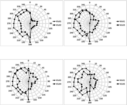
Kinetic VF was measured by Goldmann Perimetry (III4e target) before and after the prism prescribed. The patient was reminded to fixate at the fixation target during measurement. Before the use of prism, the patient had a complete right hemianopia along the vertical midline (Figure 1). With the aid of Fresnel-prism, there was a gradual expansion of temporal field and thus the overall VF became more symmetrical: an average of 10 (Visit 1) to 15 (Visit 5) degrees increase in temporal VF (across all meridians) when compared to the one in the first visit.
Figure 1 : Visual field test by Goldmann perimetry at five visits. Circle and square contours refer to the visual field measured at pre- and postinterventions respectively. Each grey grid represents 10 degrees in the measured VF. When compared the temporal field in Visit 1 and 5, there was an average increase in VF for 10 to 15 degrees.
Eye-hand coordination
Eye-hand coordination was examined by the Sports Vision Trainer (SVTTM) and total time required for responding to all the lights was measured (assessing and training eye-hand coordination). Two sets of protocols were presented, 16 or 24 randomly illuminated lights. SVT a device which is wall-mounted electronic board containing 80 touch-sensitive LED lights (10 x 8, Width x Height). Typically, lights are illuminated on the panel one at a time and the participant is required responding to the light by touching it as quickly as possible. In this study, we selected the “proaction mode”, in which lights were lit one at time and changed its location only after the patient touched that light. The completion time decreased by 35% after prism intervention (Table 2). One reason for such large improvement was the benefit of field expansion by the prism. However, a practice effect through repeated measures across visits could be another reason.
Mobility performance
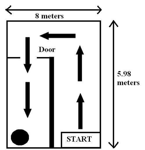
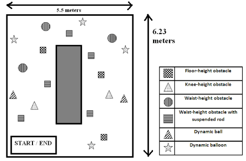
Mobility assessment was assessed by walking time and errors made for uncluttered and cluttered indoor pathways. An uncluttered pathway referred to the walkway without obvious obstacles and pedestrians (Figure 2a). A cluttered pathway referred to a mobility track, which consisted of 13 stationary (Figure 2b) obstacles and 6 dynamic obstacles*. Dynamic obstacles were only triggered when the patient crossed the sensors along the track. Although his caregiver reported significant improvement in mobility performance after prescribing the prism, objective measures could only partly agree with their subjective report. Walking speed in the uncluttered pathway was faster than that in cluttered pathway because the patient was informed that the pathway was free from obstacles other than clinic furniture and some large equipment (Table 2). After prescribing the prism, the patient walked slightly slower in both uncluttered and cluttered pathways, but made substantially fewer errors in terms of number of contacts (in particular the head-height objects) on obstacles.
Figure 2a : A schematic diagram of an uncluttered indoor walkway. The patient was requested to walk from the START to the target, which was indicated by the black circle. The length of the pathway is 19.96 meters. Arrows are only for the ease of presentation and they were not included during measurement.
Figure 2b : A schematic diagram of a cluttered indoor walkway (baseline) with four types of static (3 floor-heights, 2 knee-heights, 3 waist-heights and 5 waist-heights with extension) and two types of dynamic obstacles (2 dynamic balls and 4 dynamic balloons). The patient was required to walk through the pathway in an anti-clockwise direction (start from the left bottom corner) and avoid hitting any obstacles. The length of the pathway is 17.96 meters. Locations of objects altered for each visit to minimize the memorization of the pathway.
Self-perceived mobility performance
Patient’s self-perceived mobility performance was examined by Independent Mobility Questionnaire (IMQ) [11]. Of the 12 areas of difficulty originally reported with IMQ, improvement was noted in all areas and the most significant improvement (Moderate difficulty to No difficulty) in 9 areas is shown in Table 3. Improvement in the described activities in IMQ was also reported by the care provider, in which the increase in independence of walking up and down the stairs was obvious. Time is required to adapt to the ghostly and blurry image due to prismatic shift. Wearing schedule increased from 1 to 2 hours per day and 3 to 4 days per week during the first 6 weeks to 6 to 7 hours per day for 7 days per week afterwards. To summarize, the 10-month vision rehabilitation with the prism prescription revealed some improvement in VF, obstacles awareness during mobility, eye-hand coordination and self-independence.
    Key: 1= No difficulty;    2= mild difficulty; 3= moderate difficulty


    Question
    Visit 1
    Visit 2
    Visit 3
    Visit 4
    Visit 5


    6
    Moving about    at Stores
    3
    2
    1
    1
    1


    10
    Using public transportation
    3
    2
    1
    2
    1


    13
    Walking up steps
    3
    3
    1
    2
    1


    14
    Walking down    steps
    3
    2
    2
    3
    1


    16
    Stepping off curbs
    3
    3
    1
    2
    1


    17
    Walking through    doorways
    3
    2
    2
    1
    1


    24
    Being aware of another person’s presence?
    3
    2
    2
    1
    1


    27
    Head-height objects
    3
    2
    1
    1
    1


    28
    Shoulder-height objects
    3
    2
    2
    1
    1
Table 3: Self-evaluation on Independent Mobility Questionnaire among five visits (only the questions which showed improvement from “moderate difficulty” to “no difficulty” is shown).
Discussion
Our case study demonstrated the co-management by occupational therapists and optometrists for a patient with multiple deficits in physical, cognitive and visual perspectives due to pituitary adenoma and secondary stroke. After the rehabilitation by occupational therapists, the patient showed significant improvements in his muscular activity, cognitive function and functional performance. These improvements enhanced his self-independence during locomotion and daily activities, which might further advance the effectiveness of other types of rehabilitation. Temporal VF defect was resulted because of the compression on visual pathway (optic chiasma). After prescribing an appropriate prism for field expansion with sufficient training in vision rehabilitation, patient’s functional performance in locomotion and spatial awareness on the affected side was significantly improved. The field expansion through Fresnel prism might trigger the change in cortical representation [12,13], improvement in vision and limb activation in stroke incidence [14,15]. Despite of the substantial benefits in various aspects of functional performances through occupational and vision rehabilitation, it is plausible that our patient might achieve more efficacious interventions if a concurrent multidisciplinary rehabilitation program would have been offered. Several clinical studies have demonstrated the effectiveness of multidisciplinary vision rehabilitation [16-18]. Unfortunately, only very few healths care systems provide this type of rehabilitation for patients with stroke or brain injury. To facilitate the rehabilitation in both sensory (vision) and motor (physical movement) systems, early interventions in visual and physical function would provide better prognosis in early recovery period of more potential neural plasticity [19,20]. Clinically, many visual problems in patients with acquired brain injury may be asymptomatic or neglected, resulting in little or no management. Our recent study revealed a high prevalence of stroke-related visual problems in the post-stroke population [21]. To minimize the potential impediment to stroke rehabilitation, early recognition and intervention (e.g. vision rehabilitation) of visual problems is essential. Hence, multidisciplinary management on patients with acquired brain injury is of essence importance to provide optimal rehabilitation for improving patient’s quality of life.
Acknowledgement
We thank Prof. Peter Swann for reviewing the manuscript. This work was supported by School of Optometry, The Hong Kong Polytechnic University grants (A-PD0T).
Foot Notes:
*Static obstacles included 4 types of obstacles: three 210 mm X 297 mm grey boxes (simulating obstacles on the floor), three 297 mm X 1050 mm grey paper boxes (simulating obstacles at knee-height), three 1500 mm height chair (representing obstacles at waist-height) and 5 gray foam tubing (300 mm) suspended on 1500 mm height chair (representing obstacles at waist-height). Dynamic obstacles included 2 types of obstacles: four 300 mm diameter free-falling balloon (simulating obstacles dropped in front of the pedestrian) and 2 swinging plastics ball (simulating obstacles coming from the side).
References
- Ezzat S, Asa SL, Couldwell WT, Barr CE, Dodge WE, Vance ML, et al. The prevalence of pituitary adenomas: a systematic review. See comment in PubMed Commons below Cancer. 2004; 101: 613-619.
- Ebersold MJ, Quast LM, Laws ER Jr, Scheithauer B, Randall RV. Long-term results in transsphenoidal removal of nonfunctioning pituitary adenomas. See comment in PubMed Commons below J Neurosurg. 1986; 64: 713-719.
- Peli E. Field expansion for homonymous hemianopia by optically induced peripheral exotropia. See comment in PubMed Commons below Optom Vis Sci. 2000; 77: 453-464.
- Laws ER Jr. Vascular complications of transsphenoidal surgery. See comment in PubMed Commons below Pituitary. 1999; 2: 163-170.
- Erfurth EM, Hagmar L. Cerebrovascular disease in patients with pituitary tumors. See comment in PubMed Commons below Trends Endocrinol Metab. 2005; 16: 334-342.
- Fong KN, Chan MK, Ng PP, Tsang MH, Chow KK, Lau CW, et al. The effect of voluntary trunk rotation and half-field eye-patching for patients with unilateral neglect in stroke: a randomized controlled trial. See comment in PubMed Commons below Clin Rehabil. 2007; 21: 729-741.
- Sandford J, Browne R, Turner A. The Captain’s log cognitive training system. Computer software. Richmond, VA: Brain Train. 1996.
- Vanier M, Gauthier L, Lambert J, Pepin EP, Robillard A, Dubouloz CJ, et al. Evaluation of left visuospatial neglect: Norms and discrimination power of two tests. Neuropsychology. 1990; 4: 87-96.
- Van Deusen J. Normative data for ninety-three elderly persons on the schenkenberg line bisection test. Phys Occup Ther Geriatr. 1985; 3: 49-54.
- Schmiedecke S, Jose R. Prism therapy in low vision rehabilitation. 2005; 1282: 709-713.
- Turano KA, Geruschat DR, Stahl JW, Massof RW. Perceived visual ability for independent mobility in persons with retinitis pigmentosa. See comment in PubMed Commons below Invest Ophthalmol Vis Sci. 1999; 40: 865-877.
- Maravita A, McNeil J, Malhotra P, Greenwood R, Husain M, Driver J. Prism adaptation can improve contralesional tactile perception in neglect. See comment in PubMed Commons below Neurology. 2003; 60: 1829-1831.
- Rode G, Pisella L, Rossetti Y, Farné A, Boisson D. Bottom-up transfer of sensory-motor plasticity to recovery of spatial cognition: visuomotor adaptation and spatial neglect. See comment in PubMed Commons below Prog Brain Res. 2003; 142: 273-287.
- Harvey M, Hood B, North A, Robertson IH. The effects of visuomotor feedback training on the recovery of hemispatial neglect symptoms: assessment of a 2-week and follow-up intervention. See comment in PubMed Commons below Neuropsychologia. 2003; 41: 886-893.
- Eskes GA, Butler B, McDonald A, Harrison ER, Phillips SJ. Limb activation effects in hemispatial neglect. See comment in PubMed Commons below Arch Phys Med Rehabil. 2003; 84: 323-328.
- Hinds A, Sinclair A, Park J, Suttie A, Paterson H, Macdonald M. Impact of an interdisciplinary low vision service on the quality of life of low vision patients. See comment in PubMed Commons below Br J Ophthalmol. 2003; 87: 1391-1396.
- Lamoureux EL, Pallant JF, Pesudovs K, Rees G, Hassell JB, Keeffe JE. The effectiveness of low-vision rehabilitation on participation in daily living and quality of life. See comment in PubMed Commons below Invest Ophthalmol Vis Sci. 2007; 48: 1476-1482.
- Wang BZ, Pesudovs K, Keane MC, Daly A, Chen CS. Evaluating the effectiveness of multidisciplinary low-vision rehabilitation. See comment in PubMed Commons below Optom Vis Sci. 2012; 89: 1399-1408.
- Brainin M, Olsen TS, Chamorro A, Diener HC, Ferro J, Hennerici MG, et al. Organization of stroke care: education, referral, emergency management and imaging, stroke units and rehabilitation. European Stroke Initiative. See comment in PubMed Commons below Cerebrovasc Dis. 2004; 17: 1-14.
- Prvu Bettger JA, Stineman MG. Effectiveness of multidisciplinary rehabilitation services in postacute care: state-of-the-science. A review. See comment in PubMed Commons below Arch Phys Med Rehabil. 2007; 88: 1526-1534.
- Siong KH, Woo GC, Chan DY, Chung KY, Li LS, Cheung H, et al. Prevalence of visual problems among stroke survivors in Hong Kong Chinese patients. Clin Exp Optom. 2014; [In Press].


