
Clinical Image
Austin J Clin Ophthalmol. 2023; 10(1): 1137.
Combined Cilioretinal Artery and Branch Retinal Vein Occlusion Revealing a Carotid Artery Disease
Bouirig K*and Benkirane R
Mohammed 5 University of Rabat, Specialty Hospital of Rabat, Morocco.
*Corresponding author: Kawtar Bouirig Mohammed 5 University of Rabat, Specialty Hospital of Rabat, CHU Ibn Sina, Morocco.
Received: November 26, 2022; Accepted: January 04, 2023; Published: January 10, 2023
Clinical Image
We report the case of a healthy 55-year-old patient, who consulted at the ophthalmology emergency for a rapidly progressive unilateral visual impairment of the left right eye without neurological signs. Fundus examination of the right eye found retinal whitening following the territory of the cilioretinal artery corresponding to cilioretinal artery occlusion; dilated and tortuous retinal veins, and scattered retinal hemorrhages on the lower quadrants as well a puddle-like hemorrhages at the interpapillomacular area (Figure 1: Panel A). Fluoresceine angiography targeted a delayed perfusion of the inferior temporal branch vein (arrows) (Figure1: panel B). Macular OCT showed moderate macular thickening in the nasal macula and hyper reflectivity of the inner layers of the retina (Figure 2).
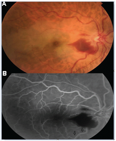
    Figure 1: (A) Fundus of the right eye: double occlusion of the cilioretinal

artery and a retinal venous branch. (B) Fluorescein angiography

showing delayed perfusion of the inferior temporal venous

branch (arrows).
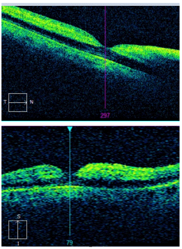
Figure 2: Macular OCT of the right eye.
The determination of ESR and CRP ruled out Horton’s disease. The cardiovascular assessment highlighted very tight carotid stenosis and high blood pressure.