Clinical Image
Austin J Clin Ophthalmol. 2023; 10(1): 1138.
Imminent Perforation of a Post-Traumatic Scleromalacia
Bouirig K*, Akkoumi A and Cherkaoui O
Mohammed 5 University of Rabat, Specialty Hospital of Rabat, Morocco.
*Corresponding author: Kawtar Bouirig Mohammed 5 University of Rabat, Specialty Hospital of Rabat, CHU Ibn Sina, Morocco.
Received: November 26, 2022; Accepted: January 09, 2023; Published: January 16, 2023
Clinical Image
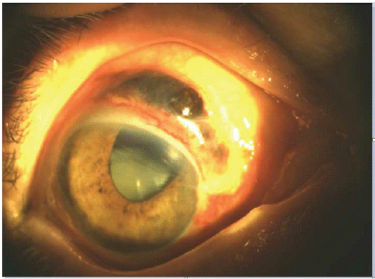
Scleromalacia is a degenerative thinning of scleral tissue. Their general causes are dominated by inflammatory rheumatism and systemic vasculitis. The traumatic cause is very rare. We report a rare case of scleromalacia following a minor trauma with localized hernia of the uveal tissue covered by an intact conjunctiva. This is a 90-year-old patient, with no particular history, who presented for consultation following the finding of the family the

presence of a pigmented mass which has been gradually increasing in size in the left eye over one month. The anamnesis revealed a recent notion of contusive trauma by elbowing and chronic constipation. The ophthalmological examination found a lesion relatively pigmented measuring 3 mm wide, parallel to the limbus going from 11 o’clock at 2 o’clock with a margin of healthy sclera, the sclera is very thin but seems perforated at 10 o’clock with herniated choroid covered by an intact conjunctiva; furthermore, the pupil is completely aspirated upwards with the presence of a dense subluxated cataract. The patient refused any surgical procedure; however, we had nevertheless requested a systemic assessment to eliminate non-traumatic causes. Our diagnosis was retained in front of the negativity of the assessment and the traumatic context.