Case Report
Austin J Clin Ophthalmol. 2023; 10(1): 1139.
Post-Partum Pituitary Apoplexy: A Case Report
Kawtar Bouirig*, Romaissae Benkirane, Abdellah Amazouzi, Nourdine Boutimzine and Lalla Ouafae Cherkaoui
Ophthalmology Department “A”, Ibn Sina University Hospital (Hôpital des Spécialités), Mohammed V University, Rabat, Morocco.
*Corresponding author: Kawtar Bouirig Ophthalmology Department “A”, Ibn Sina University Hospital (Hôpital des Spécialités), Mohammed V University, Rabat, Morocco.
Received: November 26, 2022; Accepted: January 09, 2023; Published: January 16, 2023
Abstract
Pituitary apoplexy is a potentially life threatening emergency that should be borne in mind to an early diagnosis of this extremely rare condition It is a clinical syndrome characterized by the sudden onset of headache, nausea, vomiting, visual impairment, and decreased consciousness, caused by hemorrhage and/or infarction of the pituitary gland .This article presents a case of postpartum pituitary apoplexy occurring in a 23 year old primiparous patient, this finding requires multidisciplinary approach involving expert specialists for medical or surgical treatment.
Gestational pituitary apoplexy should be suspected whenever headache and neurological disorders such as nausea and photophobia are reported during the postpartum period.
Keywords: Pituitary apoplexy; Case report; Post-partum; Headache; Visual impairment
Introduction
Pituitary apoplexy is a rare endocrine emergency. It is a clinical syndrome characterized by the sudden onset of headache, nausea, vomiting, visual impairment, and decreased consciousness, caused by hemorrhage and/or infarction of the pituitary gland. Pituitary apoplexy has very rarely been described during pregnancy and post partum period, when it is potentially lifethreatening, if unrecognized. We present the case of postpartum pituitary apoplexy revealed by unilateral visual impairment and strong headache.
Patient information
We report a case of a 23 year old patient, primiparous, with no prior medical history, was admitted to ophthalmic emergencies for sudden onset severe headache associated with nausea, a sudden vision loss of the right eye.
Clinical findings
Ocular examination showed a best corrected visual acuity at 2/10 in the right eye and 8/10 in the left eye, normal ocular motility and intraocular pressure. Anterior segment examination was unremarkable .Fundus examination revealed a pallor of the optic discs on both sides more marked in the right eye (Figure 1).

    Figure 1: Retinal photography showing pallor optic discs, marked in

the right eye (PANEL A).

    Figure 2: T1-weighted MRI scans of the brain reveal a hemorrhagic

pituitary adenoma with suprasellar extension.

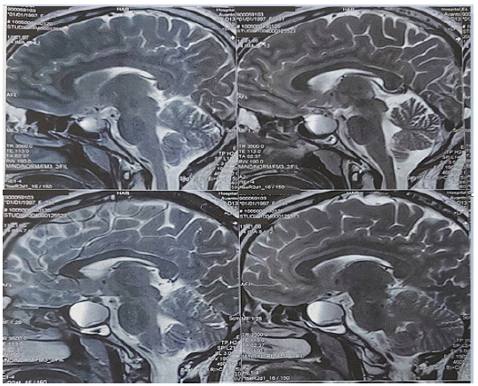
    Figure 3: T2-weighted MRI scans of the brain reveal a hemorrhagic

pituitary adenoma with suprasellar extension.

Figure 4: Visual field test objectifying bilateral quadrantanopsia.
Therapeutic intervention
Treatment with intravenous dexamethasone, thyroxin 50 μg was initiated immediately, followed by oral hydrocortisone and levo-thyroxine.
Three weeks after the apoplectic episode, the patient underwent a transethmoidal-trans- sphenoidal approach and radical tumor removal.
Follow up and outcomes.
A short-term clinical, laboratory, and instrumental follow-up was scheduled. Three months after the surgery her visual acuity and visual field. These findings were the same when reviewed one year after surgery. The patients remains clinically and neurologically stable.
Discussion
Non-functioning adenomas remains asymptomatic and can continue growing progressively, in contrary, functioning adenomas are usually detected before the apoplexy [1,2]. The case described here did not show any clinical sign of a functioning adenoma.
Pituitary apoplexy mostly occurs spontaneously [3], precipitating factors can be identified in 10-40% of cases [4] including sudden changes in blood flow within the pituitary gland, an imbalance between stimulation of the pituitary gland and increased blood flow within the pituitary adenoma, anticoagulated states [4], undergoing surgery [5] [6], endocrine stimulation tests , and post-partum [7].
Pituitary apoplexy occurs mostly in a previously unknown history of pituitary mass. The diagnosis can be challenging given its similarities with many neurological pathologies and other life-threatening
The most common features at presentation are sudden onset headache and ophthalmoplegia, followed by hypopitutarism [8,9]. The patient had visual defect due to compression of the optic chiasm are often identified due to visual impairment or other mass effect symptoms [7].
It’s a potentially life-threatening emergency that should be borne in mind to an early diagnosis of this extremely rare condition for an early management
This state can be managed by surgery or by conservative treatment. Conservative management requires immediate IV administration of high-dose glucocorticoids, hormone replacement therapy and short-term follow up.
A retrospective study of 33 cases of pituitary apoplexy has shown that in patients who present with no visual deficit or evidence of early improvement in visual deficit, a conservative (non surgical) approach is safe. Otherwise, like in our case, a surgical management is preferable [10].
Surgical treatment of pituitary adenomas usually includes both microscopic and endoscopic trans-sphenoid approaches. However, because of their extension and involvement of neurovascular structures, these tumours are associated with increased treatment morbidity and mortality, increased recurrence rates, and overall poor outcome and long-term prognosis. Careful postoperative surveillance of patients following partial removal of a pituitary adenoma is therefore mandatory. Close and effective communication between neurosurgeons, neuroophthalmologists and endocrinologists is encouraged to assure.
Conclusion
Pituitary apoplexy is a rare condition that can present with neurological signs of mass effects and corticotropic deficiency. The case reported here can be viewed as a model for a rare but life-threatening disorder that requires early diagnosis and prompt management with close multidisciplinary collaboration.
References
- Nawar RN, Mannan AD, Selman WR, Arafah BM. Pituitary tumor apoplexy: A review. J Intens Care Med. 2008; 23: 75-90.
- Dubuisson AS, Beckers A, Stevenaert A. Classical pituitary tumour apoplexy: Clinical features, management and outcomes in a series of 24 patients. Clin Neurol Neurosurg. 2007; 109: 63-70.
- Fernandez A, Karavitaki N, Wass JA. Prevalence of pituitary adenomas: A community-based, cross-sectional study in Banbury (Oxfordshire, UK). Clin Endocrinol. 2010; 72: 377-382.
- Fanous AA, Quigley EP, Chin SS, Couldwell WT. Giant necrotic pituitary apoplexy. J Clin Neurosci. 2013; 20: 1462-1464.
- Romano A, Ganau M, Zaed I, Scibilia A, Oretti G, et al. Primary endoscopic management of apoplexy in a giant pituitary adenoma. World Neurosurg. 2020; 142: 312-313.
- Goel A, Deogaonkar M, Desai K. Fatal postoperative “pituitary apoplexy”: Its cause and management. Br J Neurosurg. 1995; 9: 37-40.
- Perotti V, Dexter M. Post-partum pituitary apoplexy with bilateral third nerve palsy and bilateral carotid occlusion. Case Rep. J Clin Neurosci. 2010; 17: 1328-1330.
- Randeva HS, Schoebel J, Byrne J, Esiri M, Adams CB, et al. Classical pituitary apoplexy: Clinical features, management and outcome. Clin Endocrinol. 1999; 51: 181-188.
- Ayuk J, McGregor EJ, Mitchell RD, Gittoes NJ. Acute management of pituitary apoplexy-surgery or conservative manage-ment? Clin Endocrinol. 2004; 61: 47-752.
- John Ayuk, Elizabeth J. McGregor, Rosalind D. Mitchell, Neil JL. Gittoes. 2004.