Case Report
Austin J Clin Ophthalmol. 2023; 10(3): 1149.
Unilateral Exophthalmia Revealing Postpartum Thrombophlebitis
Debbabi Y*; Mouzari Y; Abdellaoui T; Mouzari Y; Oubaaz A
Department of Ophtalmology, Mohamed V Military Hosipital, Rabat, Morocco
*Corresponding author: Debbabi YDepartment of Ophtalmology, Mohamed V Military Hosipital, Rabat, Morocco.
Received: February 08, 2023 Accepted: March 25, 2023 Published: April 01, 2023
Abstract
Introduction: Cerebral thrombophlebitis of the peripartum is a rare entity but its occurrence is serious and can compromise the vital prognosis. The clinical picture is variable, which makes early diagnosis difficult.
Patients and Methods: We report the case of a 28-year-old patient, on oral contraception for 3 years, with no previous history; who presented rapidly progressive bilateral visual acuity loss associated with headaches. The examination revealed a painless exophamia of the left eye without motor deficit, a slight chemosis with subconjunctival venous vasodilatation, associated with a bilateral papillary edema confirmed by OCT. CT scan showing a cerebral thrombophlebitis of the superior longitudinal sinus extended to the jugular vein with slight cerebral edema. The patient was immediately admitted to the intensive care unit and put on heparin therapy and additional immunological tests.
Discussion: Pregnancy and postpartum increases the risk of thrombotic events. The most common cause in women of childbearing age is hypercoagulability, associated with the postpartum period, pregnancy, or oral contraceptives which is the case of our patient. Clinical symptoms are very polymorphic. The main thing is to think about it in order to make an early diagnosis. The positive diagnosis is neuroradiological. Improved diagnostic methods and consequent earlier treatment have markedly improved the prognosis of CVT in recent year.

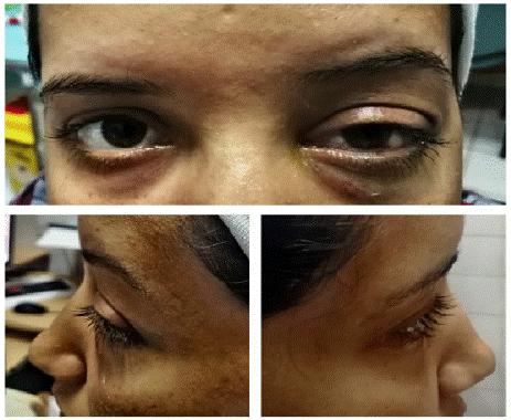
Figure 1: photo taken from the front and from the right and left sides showing the ptosis with the clinical presence of a discrete exophthalmia

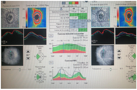
Figure 2: Papillary OCT showing the important bilateral papillary oedema.

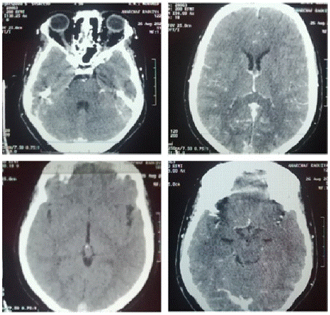
Figure 3: Spontaneous hyperdense appearance of the superior longitudinal sinus, the right lateral sinus to the sigmoid sinus and the jugular gullet realizing the rope sign with opacification defect after PDC injection and parietal enhancement realizing the empty delta sign slight associated cerebral oedema.
Conclusion: Postpartum cerebral venous thrombosis is a rare but serious pathology. The clinical diagnosis is not easy because of the clinical polymorphism, it is necessary to know how to think about it even in front of purely ophthalmological signs and to ask for neuro-radiological examinations to confirm it. The prognosis remains good if the diagnosis is made in time and if the treatment is started early.
Introduction
Cerebral thrombophlebitis of the peripartum is a rare entity but its occurrence is serious and can compromise the vital prognosis. It represents 10 to 20% of CVTs. Pregnancy and postpartum are situations at risk because of the physiological hypercoagulability that accompanies them. The clinical picture is variable, which makes early diagnosis difficult.
Patients and Methods
We report the case of a 28-year-old patient, on oral contraception for 3 years, with no previous history, G1P1EV0, who gave birth to a baby 15 days ago for severe pre-eclampsia complicated by retroplacental hematoma with fetal death in utero, and who presented for 4 days with an HTIC syndrome without sensory-motor deficits, vigilance disorders or seizures, without fever She was admitted to our unit for a rapidly progressive bilateral visual acuity loss associated with headaches.
The examination revealed a painless exophamia with ocular ptosis of the left eye without motor deficit, a slight chemosis with subconjunctival venous vasodilatation, associated with a bilateral papillary edema confirmed by OCT.
In front of this picture, a CT scan was urgently requested, showing a cerebral trhombophlebitis of the superior longitudinal sinus extended to the jugular vein with slight cerebral edema.
The patient was immediately admitted to the intensive care unit and put on heparin therapy and additional immunological tests.
Discussion
Pregnancy and postpartum increases the risk of thrombotic events (hypercoagulability). The average incidence of postpartum cerebral thrombophlebitis is 12 cases per 100,000 deliveries [1]. They usually occur early after de
The most common cause in women of child bearing age is hypercoagulability, associated with the postpartum period, pregnancy, or oral contraceptives. In a consecutive study by Bousser et al, oral contraception was noted as the only possible etiology of CVT in 14 of 135 patients (10%), and in the ISCVT study, 46% of women with CVT were on oral contraceptives. [2]
However, a recent study seems to demonstrate the increased risk of venous thromboembolic events, also between 6 and 12 weeks postpartum [2].
The clinical symptomatology of these CPTs is very polymorphic. The main thing is to think about it in order to make an early diagnosis. Their onset may be early (less than 48 hours after delivery) in 28% of cases, subacute (between 48 hours and 30 days) in 47% of cases or late (more than 30 days) in 25% of cases [3,4]. CPT should be suspected when the parturient develops symptoms associating to varying degrees intracranial hypertension (headache, vomiting, papilledema, consciousness disorders) and/or focal neurological deficit and/or seizures [5].
The positive diagnosis can only be neuroradiological [6]. Cerebral CT without and with contrast injection is still performed as a first line of treatment. It remains normal in 4 to 25% of patients with CPT [5]. The typical appearance is the presence of a delta sign, found in about 25% of cases. It appears as a hypodense area surrounded by contrast. Another direct sign is the fresh thrombus, which appears as a spontaneous hyperdensity at the site of the thrombosed vein [7]. Indirect signs visible on the cerebral CT scan are essentially venous infarcts but also the existence of cerebral edema.
In the acute phase patients with CVT are treated by intravenous heparin or low molecular weight heparins in therapeutic dosage. There after the use of an oral anticoagulant (target INR 2.0–3.0) for a limited period of 3–12 months is recommended.
Improved diagnostic methods and consequent earlier treatment have markedly improved the prognosis of CVT in recent year [8].
Conclusion
Postpartum cerebral venous thrombosis is a rare but serious pathology. The clinical diagnosis is not easy because of the clinical polymorphism, it is necessary to know how to think about it even in front of purely ophthalmological signs and to ask for neuro-radiological examinations to confirm it. The prognosis remains good if the diagnosis is made in time and if the treatment is started early
References
- Saposnik G, Barinagarrementeria F, Brown RD, Bushnell CD, Cucchiara B, et al. Diagnosis and management of cerebral venous thrombosis: a statement for healthcare professionals from the American Heart Association/ American Stroke Association. Stroke. 2011; 42: 1158–92.
- Kamel H, Navi BB, Sriram N, Hovsepian DA, Devereux RB, et al. Risk of thrombotic event after the 6-week postpartum period. N Engl J Med. 2014; 370: 1307–15.
- Aissi M, Boughammoura-Bouatay A, Frih-Ayed M. Thrombophlébite cérébrale au cours de la grossesse. Gynécologie Obstétrique & Fertilité. 2016; 44: 129–131.
- Ameri A, Bousser MG. Cerebral venous thrombosis. Neurol Clin. 1992; 10: 87–111.
- Shah M, Agarwal N, Gala NB, Prestigiacomo CJ, Gandhi CD. Management of dural venous sinus thrombosis in pregnancy. European Journal of Vascular and Endovascular Surgery. 2014; 48: 482–484.
- Arquizan C. Thrombophlébites cérébrales: aspects cliniques, diagnostic et traitement. Réanimation. 2001; 10: 383–92.
- Wendling LR. Intracranial venous sinus thrombosis: diagnosis suggested by computed tomography. AJR. 1978; 130: 978–80.
- Urs Fischer, Krassen Nedeltchev, Jan Gralla, Caspar Brekenfeld, Marcel Arnold Thromboses veineuses cérébrales:mise à jour ¬ Hôpital de l’Ile, Université de Berne a Clinique neurologique et Policlinique, b Institut de neuroradiologie diagnostique et interventionnell.