Review Article
Austin J Clin Ophthalmol. 2023; 10(4): 1151.
Regener-Eyes® Ophthalmic Solution: A New Therapeutic Agent to Relieve Dryness of the Eye for Tear Hyperosmolarity-Induced Pathological Changes in the Eyes of Patients Suffering From Dry Eye Discomfort
Carl Randall Harrell*
Regenerative Processing Plant, LLC, United States of America
*Corresponding author: Carl Randall Harrell Regenerative Processing Plant, LLC, 34176 US Highway 19 N, Palm Harbor, FL34684, Florida, USA. Tel/Fax+1 800-781-0818 Email: dr.harrell@regenerativeplant.org
Received: February 28, 2023 Accepted: March 29, 2023 Published: April 05, 2023
Abstract
Tear hyperosmolarity is an initial and crucial step in the development, progression and aggravation of dry eye discomfort. Decreased tear secretion or altered tear composition leads to tear film instability/imbalance which in Dry Eye (DE) patients results in abnormally rapid breakup of the tear film after blinking. Numerous structural changes in epithelial cells and mucin-producing goblet cells develop as a consequence of exposition of these cells to the hyperosmolar tears. Tear hyperosmolarity causes oxidative stress, disruption of DNA repair system and induces DNA damage in the cells of ocular surface and lacrimal system, leading to their apoptosis. An injury of lacrimal glands results in decreased tear secretion, enabling the creation of “positive loop” that leads to the DE progression and aggravation. Accordingly, eye drops, which alleviate tear hyperosmolarity and restore tear homeostasis at corneal surface, will break up vicious DEcycle and will relieve eye pain, irritation, discomfort, and vision disturbance in DE patients. Regener-Eyes® Ophthalmic Solution is a hypotonic solution enriched with osmoprotectants (that address the hyperosmolarity of a tear film), which helps support tear stability and contribute to relieve dryness of the eye in DE patients. In this article, we summarized current knowledge and future perspectives about topical administration of Regener-Eyes® Ophthalmic Solution to relieve dryness of the eye in DE patients.
Keywords: Tear hyperosmolarity; Eye drops; Eye inflammation; Therapy; Dry eye
Introduction
Dry Eye (DE) disease, also known as keratoconjunctivitis sicca or dysfunctional tear syndrome, is a common, multifactorial disease of the lacrimal system and ocular surface characterized by a deficiency in quality and/or quantity of the tear fluid [1].
Since the ocular surface is highly exposed to environmental hazards, efficient tear production and optimal tear turnover is essential for appropriate eye function [2]. The tear film, lacrimal glands (main and accessory lacrimal glands), meibomian glands (oil-producing glands positioned along the edge of the eyelids that create oily layer outside of the tear film, keeping tears from drying up too quickly), mucin-producing goblet cells, ocular surface secretory cells, lacrimal outflow pathways, corneal and conjunctival epithelial cells work together and function as a Lacrimal functional Unit (LFU) to maintain the tear film, protect the transparency of the cornea and the integrity of the ocular surface [3]. Importantly, the LFU is not an isolated system, and it functions in association with nervous and endocrine systems. Damage of any LFU component and/or development of neural and endocrine disease (the dysfunction of sensory and motor nerves, hormone imbalance) will destabilize the tear film and will lead to the development of DE [1,3]. An unbalanced tear film is not able to provide sufficient nourishment or protection to the ocular surface and, therefore, usually results in permanent damage of the corneal nerve fibers, corneal and conjunctival epithelial cells [1,3].
Dry eye disease is often classified into two primary subtypes: Aqueous Tear-Deficient Dry Eye (ADDE), characterized by inefficiency or inability of the lacrimal glands to produce tears, and Evaporative Dry Eye (EDE), typically attributed to excessive evaporation of the tear fluid [4]. ADDE may have an autoimmune origin or is attributed to a compromise in the LFU integrity [4]. EDE is the more common form of dry eye disease and is frequently associated with Meibomian Gland Dysfunction (MGD) characterized by modification or reduction of tear fluid lipids, due to which, integrity and quality of the tear fluid may be compromised [4]. Although traditionally, dry eye disease has been classified into these two subtypes, there is considerable overlap between them. As such, dry eye disease is the most often characterized as a "hybrid" or "mixed" form of these two subtypes, where in each subtype adopts some clinical features of the other, initiating and exacerbating its pathology [4].
Multifactorial nature of dry eye disease involves several inter-related underlying pathologies, including the loss of homeostasis, chronic eye inflammation, instability and hyperosmolarity of the tears which leads to the neurosensory dysfunction and visual disturbance [1,5]. These detrimental events create a “pathological loop" which promotes progression and aggravation of dry eye disease [1]. DE is usually manifested by dryness, grittiness, scratchiness, soreness, irritation, burning, watering, foreign body sensation, eye fatigue and reduced functional visual acuity [5,6]. Since significantly impaired performance of vision-dependent daily activities diminishes quality of life of dry eye disease patients, better understanding of pathological steps in dry eye disease pathogenesis is of crucial importance for appropriate dry eye disease treatment [1,6].
Tear Hyperosmolarity: An Initial and Crucial Step in the Development, Progression and Aggravation of Dry Eye Disease
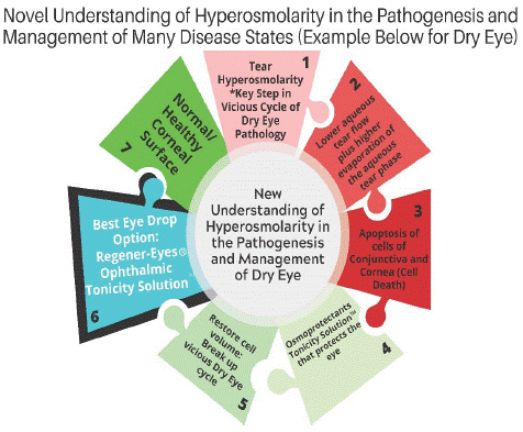
Decreased tear secretion or altered tear composition leads to tear film instability/imbalance which in dry eye disease patients results in abnormally rapid breakup of the tear film after blinking [7]. This leads to local drying and hyperosmolarity of the exposed surface which provokes cell death, injury of LFU components and induces a cascade of detrimental inflammatory events, resulting in eye pain, irritation, discomfort, and vision disturbance in dry eye disease patients (Figure 1) [4,8].

Figure 1: Tear hyperosmolarity: an initial and crucial step in the development, progression and aggravation of DE. Decreased tear secretion or altered tear composition leads to the tear film instability/imbalance which lead stolocal drying and hyperosmolarity of the exposed surface. Tear hyperosmolarity induces activation of apoptosis-related signaling pathways and causes programmed cell death of epithelial cells at corneal surface and in the lacrimal and meibomian glands. An injury of lacrimal glands results in decreased tear secretion, enabling the creation of a “positive loop” that leads to the DE progression and aggravation. Eye drops, which alleviate tear hyperosmolarity and restore tear homeostasis at corneal surface, will break up vicious DE cycle and may relieve eye pain, irritation, discomfort, and vision disturbance in DE patients. Regener-Eyes® Ophthalmic Solution is a hypotonic solution enriched with osmoprotectants which relieve dryness of the eye in DE patients.
Tear osmolarity (measured from the lower meniscus) ranges from approximately 300 to 310mOsM/kg in normal eyes [4,8]. However, dry eye disease patients, with or without tear volume reduction, have higher evaporation rates than healthy subjects, which finally results in tear hypertonicity. When osmolarity of the tears exceeds that of the epithelial cells, mechanisms of osmoregulation which protect ocular surface epithelia and preserve normal vision, are disturbed. The current cutoff for a diagnosis of DE is 316mOsM/kg, although tear hyperosmolarity in individual dry eye disease patients may reach as high as 360mOsM/kg. Tear osmolarity may be used in dry eye disease diagnostic testing. A cut-off of 316mOsM/L identifies dry eye disease more accurately than other single tests, including the Schirmer test, rose Bengal staining, and lactate levels [4,8].
Numerous structural changes in epithelial cells and mucin-producing goblet cells develop as a consequence of exposition of these cells to the hyperosmolar tears [7]. Tear hyperosmolarity causes oxidative stress, disruption of DNA repair system and induces DNA damage in the cells of ocular surface and lacrimal system, leading to their apoptosis [7]. Accordingly, epithelial and goblet cells of DE patients, which are constantly exposed to hyperosmolar tears, lose their phenotype and function. Consequently, tear hyperosmolarity reduces functional visual acuity and significantly impairs performance of vision- dependent daily activities (reading, writing, driving) of DE patients, constantly reducing their quality of life [7].
Symptoms like dryness, grittiness, scratchiness, soreness, irritation, burning, watering, foreign body sensation and eye fatigue, frequently observed in DE patients, are directly or indirectly caused by tear hyperosmolarity [1,4,7,8]. In addition to the direct injury of corneal epithelial cells and mucin-producing goblet cells, tear hyperosmolarity can indirectly induce injury of these cells by activating an inflammatory cascade in the eyes of dry eye disease patients [4,7,8]. Exposure to hyperosmotic stress activates two Mitogen-Activated Protein Kinase (MAPK) signaling pathways (C-Jun N- Terminal Kinase (JNK) and Extracellular-Regulated Kinase (ERK) pathways), resulting in an increased expression and production of pro-inflammatory cytokines. Interleukin 1 (IL-1) and Tumor Necrosis Factor Alpha (TNF-a). IL-1 and TNF- a induce enhanced expression of adhesion molecules on endothelial cells enabling a massive influx of antigen-presenting Dendritic Cells (DCs), macrophages and circulating lymphocytes in lacrimal glands and ocular surface of DE patients [7-9]. These inflammatory cells produce massive amounts of lL-1 and TNF- a and creates a "positive inflammatory loop" in the eyes of DE patients that results in the progression and aggravation of DE [7-9]. Additionally, hyperosmolarity enhances the interaction between antigen- presenting cells (DCs and macrophages) and inflammatory Thl and Thl7 effector cells that play a crucially important pathogenic role in DE progression [1,9]. DCs activate T lymphocytes in the eyes of DE patients by presenting antigens within Major Histocompatibility Complex (MHC) molecules [1]. Even minor increase in tear osmolarity may be sufficient to generate an inflammatory response in DE patients since hyperosmolarity significantly increases expression of MHC molecules on DCs, resulting in the generation of strong detrimental Th1/Thl7 cell-driven inflammation in the eyes of dry eye disease patients [7,9].
In order to remove patients from the cycle of interactions that can amplify the severity of dry eye disease, central mechanisms such as tear hyperosmolarity must be addressed [1,2,7]. Traditional approaches to correcting hyperosmolarity in dry eye disease include use of hypotonic tear substitutes which have limited persistence in the eyes, and this means that after instillation, osmolarity returns to a hyperosmolar range within approximately 1-2 minutes [1,2,7]. DE treatments may benefit from inclusion of osmoprotectants, naturally occurring compatible solutes, such as natural water, that are internalized by cells, restoring cell volume and stabilizing proteins [7]. The osmoprotective effect depends on how much osmoprotectants the cell takes up and how long it is retained. For example, glycerol can rapidly and easily enter the cell via the water channel, but also leaves the cells very quickly and, therefore, its therapeutic effects could be improved if it is combined with a protectant that acts over a longer term [1,2,7].
In line with these findings, with greater understanding of hyperosmolarity in DE development and progression, eye care professionals must consider topical administration of eye-drops which are designed specifically to address hyperosmolarity in the eyes of DE patients for longer periods of time [7].
Regener-Eyes® Ophthalmic Solution: A New Eye Drop to Relieve Dryness of the Eye that can Solution, Address Tear Hyperosmolarity
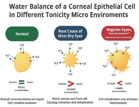
Regener-Eyes® Ophthalmic Solution is manufactured under Current Good Manufacturing Practices (cGMP), regulated by the Food and Drug Administration (FDA). Regener-Eyes® Ophthalmic Solution is a hypotonic solution, which helps alleviate discomfort, supports tear stability and contributes to helping relieve dryness of the eye. Tear hyperosmolarity is the principal step in the vicious cycle of DE and is considered the main reason for the development, progression, and aggravation of DE [4,8]. It is a state in which the osmolarity of the tear exceeds that of the epithelial cells, leading to reduced cell volume and increased concentration of solutes [4,8]. During the progression of DE, water moves out from cell causing crenation and dehydration. Regener-Eyes® Ophthalmic Solution helps to relieve dryness of the eye by possibly helping to increase hydration to the cornea, thereby helping to restore homeostasis to the corneal surface (Figure 2).

    Figure 2: Regener-Eyes® Ophthalmic Solution alleviates tear hyperosmolarity and can help restore homeostasis at corneal surface.

Tear hyperosmolarity, a state in which the osmolarity of the tear exceeds that of the epithelial cells, leads to the increased concentration of solutes. Accordingly, water moves out from cells causing crenation and dehydration. Hypotonic solution of Regener-Eyes® Ophthalmic Solution relieves dryness of the eye in DE patients.
Accordingly, when you have homeostasis of the cornea it addresses all hyperosmolarity-related issues and may prevent the development of pathological events which are elicited by exposition of ocular surface to the hyperosmolar tears. Precisely, addressing these issues in DE patients may: (i) allow hypotonic natural tear stability; (ii) prevent oxidative stress in meibomian glands, epithelial and mucin-producing goblet cells, (iii) prevent DNA damage and apoptosis of the cells at ocular surface, lacrimal system and meibomian glands enabling their better functioning; (iv) prevent hyperosmolarity-induced production of pro-inflammatory cytokines in eye-infiltrated immune cells; (v) attenuate on-going eye inflammation (by inhibiting IL-1 and TNF-a-driven inflammation in interleukin 1 Receptor antagonist (IL-1Ra) and soluble receptor of tumor necrosis factor alpha (sTNFR)-dependent manner); (vi) suppress Th1/Th17 cell-dependent detrimental immune response (by down-regulating hyperosmolarity-induced enhanced expression of MHC molecules on the membrane of DCs); (vii) enable DE patients to relieve dryness of the eye; (viii) enable improvement of visual acuity and better performance of vision-dependent daily activities.
Evidence of Regener-Eyes® Ophthalmic Solution-dependent attenuation of DE-related pathology
Therapeutic potential of Regener-Eyes® Ophthalmic Solution in the suppression of hyperosmolarity-induced eye pathology was confirmed in clinical settings. Regener-Eyes® Ophthalmic Solution efficiently alleviated ocular discomfort and pain in 131 DE patients (27 males and 104 females with a median age of 62 years (range 19-85), Regener-Eyes® Ophthalmic Solution remarkably improved all DE-related symptoms including pain, dryness, grittiness, scratchiness, soreness, irritation, burning, watering, and eye fatigue.
Significantly reduced DE questionnaires shows Visual Analogue pain Score (VAS) and Standard Patient Evaluation of Eye Dryness Questionnaire (SPEED) scores were noticed in Regener-Eyes® Ophthalmic Solution-treated patients. Regener-Eyes® Ophthalmic Solution dependent possible beneficial effects have been noticed during the entire observational period and significantly increased during the last 6 months of the follow-up. Significant decrease in VAS and SPEED scores in Regener-Eyes® Ophthalmic Solution treated patients was documented 3 months after which may indicate the beneficial effects of Regener-Eyes® Ophthalmic Solution in alleviation of ocular symptoms in patients.
Beneficial effects of Regener-Eyes® Ophthalmic Solution in the treatment of MGD-related were also demonstrated. Regener-Eyes® Ophthalmic Solution efficiently attenuated related symptoms in patients suffering from MGD. Before topical application of Regener-Eyes® Ophthalmic Solution, meibomian ducts of MGD patients were dilated while meibomian glands were enlarged and tortuous with abnormal structure.
The morphology of meibomian glands was significantly improved after 3 weeks of Regener-Eyes® Ophthalmic Solution showing the hypoilluminescent grape-like clusters. Similarly, hypoilluminescent ducts and underlying tarsus indicated beneficial effects of Regener-Eyes® Ophthalmic Solution in restoring meibomian gland and ducts morphology. Before topical application of, MGD patients reported foreign body sensation and pain in the eyes, which were accompanied with grittiness, soreness, irritation, and burning and eye fatigue.
Importantly, none of these DE-related symptoms were reported by MGD patients after 3 weeks of Regener-Eyes® Ophthalmic Solution based therapy. Significantly improved Tear film Breakup Time (TBUT) was noticed 3 weeks after Regener-Eyes® Ophthalmic Solution based treatment, which may indicate better meibomian gland function.
Significantly improved visual acuity, ocular pain relief and healing of corneal epithelial defects were also noticed in Regener-Eyes® Ophthalmic Solution-treated patients with DE which developed because of underlying autoimmune disease, Sjogren's syndrome. Four weeks of Regener-Eyes® Ophthalmic Solution also showed remarkably improved visual acuity and significantly decreased ocular pain in a 26-year-old female who suffered from severe DE and epithelial basement membrane dystrophy with recurrent corneal erosion syndrome. Similarly, 15 days of Regener-Eyes® Ophthalmic Solution-based therapy significantly alleviated neurotrophic keratitis in an 80-year-old patient. She used Regener-Eyes® Ophthalmic Solution 3-4 times/day for 4 days and 2-3 times/day for 11 days and keratitis was nearly resolved.
Safety of Regener-Eyes® Ophthalmic Solution showed in dry eye patients
Regener-Eyes® Ophthalmic Solution was well tolerated in all clinical outcomes and has an excellent safety profile. None of 131 Regener-Eyes® Ophthalmic Solution-treated patients reported any severe side effects related to the Regener-Eyes® Ophthalmic Solution, suggesting that topical application of Regener-Eyes® Ophthalmic Solution is a safe and effective therapeutic approach.
Similarly, symptoms related to recurrent corneal erosion syndrome were not observed in Regener-Eyes®Ophthalmic Solution patients during a follow-up of four months, suggesting possible beneficial effects of Regener-Eyes® Ophthalmic Solution in addressing the injured corneal epithelial cells.
Comparison Between Therapeutic Potential of Regener-Eyes® Ophthalmic Solution and Other Commercially Available Eye-drops to Relieve Dryness of the eye
One competing dry eye company has a hypertonic ophthalmic solution in which the main active ingredient is sodium chloride. The main mechanism of action is to remove water out of a swollen cornea [7]. Opposite to this approach which, as hypertonic solution, may aggravate tear hyperosmolarity-induced injury and inflammation in the eyes of DE patients, Regener-Eyes® Ophthalmic Solution is a hypotonic solution that may attenuate tear hyperosmolarity-induced pathological events.
Another competing dry eye product is a therapeutic agent that lubricates the eyes since it is anisotonic demulcent eye drop whose active ingredient is methylcellulose [1,7]. The other competing dry eye product offers an isotonic emollient eye ointment since its active ingredientis mineral oil [1,7]. These products lubricate the dry eyes of DE patients relieving dryness and irritation caused by reduced tear flow. However, the majority of tear hyperosmolarity-induced changes in phenotype and function of corneal epithelial cells and mucin-producing goblet cells could not be completely restored by these products. In contrast to these products, Regener-Eyes® Ophthalmic Solution is a hypotonic solution which helps to relieve dryness of the eye by addressing hyperosmolarity-induced apoptosis of the cells in the lacrimal system, and which may induce enhanced repair of epithelial barrier at the ocular surface of DE patients. Therefore, opposite to these other dry eye products which could only temporarily relieve dryness and irritation caused by reduced tear flow, beneficial effects of Regener-Eyes® Ophthalmic Solution can be long-lasting for patients.
Another eye drop is an isotonic vasoconstrictor eye drop whose main mechanism of action is reduction of blood vessel size, resulting in significant reduction of redness in the eyes of DE patients [1,7]. Similar to this product, Regener-Eyes® Ophthalmic Solution is also able to help by improvement in the overall microenvironment. Opposite to other dry eye drops, which are isotonic solutions, Regener-Eyes® Ophthalmic Solution is a hypotonic ophthalmic solution that may address tear hyperosmolarity-induced pathological events in the eye. Accordingly, it is not expected that these other competing dry eye drops could address hyperosmolarity-induced cell injury and eye inflammation, while beneficial effects of Regener-Eyes® Ophthalmic Solution may improve the epithelial barrier at ocular surface and on IL-lRa, sTNFR-dependent suppression of eye inflammation by improving the microenvironment in DE patients.
Conclusions
Dry eye disease is the most common corneal surface disease globally. Successfully treating these dry eye conditions, especially the severe dry eye disease that accompanies neurotropic keratitis and Sjogren’s syndrome, requires a new paradigm in our understanding of the microenvironment of the cornea. Immunosuppressive cyclosporine and treatments with recombinant protein therapy, like such as a single human nerve growth factor would not be expected to significantly change the corneal microenvironment.
New understanding of the root cause of dry eye disease before the biologic changes occur gives an early window to help address further dry eye problems from progressing. Topical administration of Regener-Eyes® Ophthalmic Solution addresses hyperosmolarity in dry eye patients. Regener-Eyes® Ophthalmic Solution can improve the microenvironment to relieve dryness of the eye. Regener-Eyes® Ophthalmic Solution may be considered as a new safe and effective topical agent in addressing dryness of the eye in patients.
Abbreviations
DE: Dry Eye; LFU: Lacrimal Functional Unit; ADDE: Aqueous Tear-Deficient Dry Eye; EDE: Evaporative Dry Eye; MGD: Meibomian Gland Dysfunction; MAPK: Mitogen-Activated Protein Kinase; JNK: C-Jun N-Terminal Kinase; ERK: Extracellular-Regulated Kinase; IL-1: Interleukin 1; TNF-a: Tumor Necrosis Factor Alpha: DCs: Dendritic Cells; MHC: Major Histocompatibility Complex; cGMP: Current Good Manufacturing Practices; FDA: Food and Drug Administration; RPP: Regenerative Processing Plant’s; IL-1Ra: Interleukin 1 Receptor Antagonist; sTNFR: Soluble Receptor of Tumor Necrosis Factor Alpha; VAS: Visual Analogue Pain Score; SPEED: Standard Patient Evaluation of Eye Dryness Questionnaire; TBUT: Tear Film Breakup Time
References
- Craig JP, Nichols KK, Akpek EK, Caffery B, Dua HS, et al. TFOS DEWS II Definition and Classification Report. Ocul Surf. 2017; 15: 276-283.
- Koh S, Rao SK, Srinivas SP, Tong L, Young AL. Evaluation of ocular surface and tear function-A review of current approaches for dry eye. Indian J Ophthalmol. 2022; 70: 1883-1891.
- Conrady CD, Joos ZP, Patel BC. Review: The Lacrimal Gland and Its Role in Dry Eye. J Ophthalmol. 2016; 2016: 7542929.
- Agarwal P, Craig JP, Rupenthal ID. Formulation Considerations for the Management of Dry Eye Disease. Pharmaceutics. 2021; 13: 207.
- Milner MS, Beckman KA, Luchs JI, Allen QB, Awdeh RM, et al. Dysfunctional tear syndrome: dry eye disease and associated tearfilm disorders -new strategies for diagnosis and treatment. Curr Opin Ophthalmol. 2017; 27: 3-47.
- Pflugfelder SC, de Paiva CS. The Pathophysiology of Dry Eye Disease: What We Know and Future Directions for Research. Ophthalmology. 2017; 124: S4-S13.
- Baudouin C, Aragona P, Messmer EM, Tomlinson A, Calonge M, et al. Role of hyperosmolarity in the pathogenesis and management of dry eye disease: proceedings of the OCEAN group meeting. Ocul Surf. 2013; 11: 246-58.
- Liu H, Begley C, Chen M, Bradley A, Bonanno J, et al. A link between tear instability and hyperosmolarity in dry eye. Invest Ophthalmol Vis Sci. 2009; 50: 3671-9.
- Li DQ, Luo L, Chen Z, Kim HS, Song XJ, et al. JNK and ERK MAP kinases mediate induction of IL-1beta, TNF-alpha and IL-8 following hyperosmolar stress in human limbal epithelial cells. Exp Eye Res. 2006; 82: 588-96.