Case Report
Austin J Clin Ophthalmol. 2023; 10(5): 1159.
Bilateral Orbital Myositis: A Rare Case Report
M Yousfi*; R Chahir; G Daghouj; L Elmaaloum; B Allali; A Elkettani
Department of Pediatric Ophthalmology, Hospital August 20, 1953, Morocco
*Corresponding author: Manal Yousfi Department of Pediatric Ophthalmology, Hospital August 20, 1953, Morocco. Email: manal.yousfi@gmail.com
Received: June 02, 2023 Accepted: June 22, 2023 Published: June 29, 2023
Abstract
The orbital idiopathic myositis is included in the group of orbital inflammatory pseudotumors. It’s an isolated inflammation, unspecific of orbital muscles. This pathology is rare in Children and it is a source of real diagnosis problems. We report the case of a 10-year-old boy who was hospitalized for bilateral painful exophthalmos. We report the case of a 10-year-old boy who was hospitalized for bilateral exophthalmos of abrupt onset, painful, axial, and non reducible with complete ophthalmoplegia of both eyes. The examination showed a retained Visual Acuity (VA) of 10/10 with a normal Fundus Examination (FO) in both eyes. The inflammatory and thyroid work-up are negative. The orbital tomodensitometry revealed a grade I exophtalmitis and a thickening of external and inferior muscles. The corticotherapy leaded to the decline of exophtalmitis andnormalization of eye movements after 1 month of treatment. The etiologic survey was negative, then the diagnosis of idiopathic myositis was kept. The orbital idiopathic myositis stills an eliminating diagnosis. Faced to a child’s exophtalmitis, other differential diagnosis must be excluded by an advanced etiologic survey.
Introduction
Idiopathic Orbital Myositis (IOM) is an inflammatory pathology affecting the oculomotor muscles. This condition is a subgroup of the inflammatory orbitopathies inflammatory orbitopathies, which include any inflammation of the orbit whose etiology is negative. It is a pathology that is rarely described in described in children, although it often poses problems of differential diagnosis with malignant tumors especially in their acute form. Through this observation and the data of the literature, we will try to literature, we will try to identify the mechanisms and the clinical and para clinical, evolutionary and therapeutic particularities of MOI. We report the case of a 10-year-old child who presented with bilateral orbital myositis revealed by a painful exophthalmos associated with ptosis with good functional recovery under corticosteroid therapy
Observation
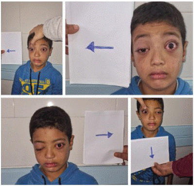
We report the case of the child A.M., aged 10 years, without any notable pathological history, who presented to the emergency room for a painful bilateral exophthalmos of sudden onset, associated with visual discomfort (Figure 1).

Figure 1: Complete ophthalmoplegia.
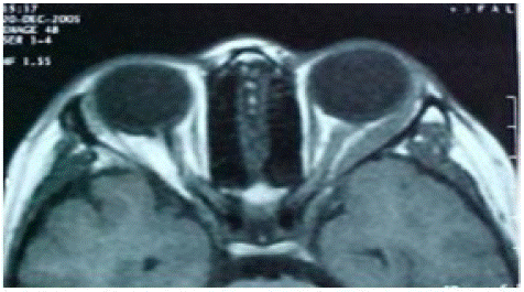
The ophthalmological examination revealed an axial exophthalmos, non reducible, associated with a ptosis of the right eye and bilateral local inflammatory signs such as conjunctival hyperhemia and palpebral edema. Oculomotor examination revealed a total limitation of oculomotor function in both eyes. VA was 10/10 in both eyes, anterior segment and FO examination were unremarkable in both eyes. An orbital ultrasound showed thickening of the oculomotor muscles, and a CT scan confirmed the presence of grade I exophthalmos with diffuse infiltration of the left lateral and inferior rectus muscles (Figure 2).

Figure 2: Diffuse infiltration of the right.
The inflammatory and infectious biological workup came back normal, as well as the thyroid workup. The diagnosis of idiopathic myositis was strongly suspected and the child was put on a bolus of solumedrol for 3 days in a row, followed by an oral dose of 1mg/Kg/d. The spectacular evolution of the exophthalmos and inflammatory signs with regression from the 2nd day of bolus day of bolus (Figure 3).

Figure 3: Full functional recovery.
Discussion
Idiopathic inflammatory myositis accounts for 10% of idiopathic orbital pseudotumors, and are more frequent in children. The etiopathogeny of this condition remains poorly known, but the autoimmune theory is the most likely [1]. The clinical manifestations of myositis are palpebral edema, chemosis, diplopia, pain and limitation of movement. Pain and limitation of movement during the contraction of the contraction of the affected muscle. All these symptomatology is characterized by a sudden onset [3]. The most affected muscles are the rectus muscles, although the although the participation of the oblique muscles has been reported. Muscle involvement is usually unilateral, but may be bilateral unilateral, but may be bilateral. The diagnosis of acute orbital myositis is based on the sudden onset of painful exophthalmos painful exophthalmos, restriction of eye movements leading to diplopia, the presence of inconstant inflammatory signs and rapid response to corticosteroid therapy [4]. In our case, the exophthalmos was of abrupt onset, associated with limited abduction of the left eye, without biological inflammatory signs. B-mode ultrasonography showed muscle thickening. CT is useful for the positive diagnosis and to eliminate other differential diagnoses. On the other hand, in dysthyroid orbitopathy, the muscles are thickened especially in their medial part sparing the tendons. A muscle biopsy is required whenever there is a there is a diagnostic doubt, or that the response to to treatment is delayed, in order to eliminate an intra-orbital tumor process, that could endanger the patient's vital prognosis. At theanatomical examination, the oculomotor muscles normally contain lymphocytes distributed in a diffuse and perivascular manner. In presence of myositis, the biopsy shows muscles infiltrated by a large number of lymphocytes and plasma cells. The muscle fibers are swollen and separated by edema and fibrosis [2]. The differential diagnoses to be considered in the presence of exophthalmos in children are rhabdomyosarcoma, lymphoma, orbital metastases, dysthyroid orbitopathy, sarcoidosis and infectious diseases (toxoplasmosis, tuberculosis, toxocariasis, etc.)[5]. The treatment of idiopathic orbital myositis is essentially based on high-dose corticosteroid therapy (1 to 1.5mg/Kg/J of prednisone) for 15 days to 1 month, followed by a decreasing maintenance dose over several months. The clinical response is so rapid that some authors have considered it as a diagnostic criterion (the American school). In our case the evolution was spectacular with no recurrences after a current follow-up of 10 months.
Conclusion
Idiopathic myositis of the orbit remains a diagnosis of elimination. It is a benign pathology for which the clinical and para-clinical assessment must be carried out as a matter of urgency in order to eliminate a possible rhabdomyosarcoma which would rhabdomyosarcoma which would require urgent treatment, and not to not to delay the start of corticosteroid therapy which is the guarantee of a cure without sequelae. In case of delay or lack of response to corticosteroid treatment, a biopsy is necessary.
References
- Souhail H, Elmoussaif H, Mouhcine Z. La myosite idiopathique de l’orbite de l’enfant à propos d’un cas. Réflexions Ophtalmologiques. 2003; 8: 31-32.
- George JL. Oculomotor disturbances due to idiopathic inflammatory orbital pseudotumor. Orbit. 1989; 8: 117-122.
- Bouhamida K, Gicquel JJ, Mercie M, Boissonnot M, Dighiero P. Diagnostic de myosite orbitaire idiopathique, à propos de 3 cas. J Fr Ophtalmol. 2002; 25: 95-96.
- Trakel SL, Hilas K. Recognition and differential diagnosis of enlarged extraocular muscles in computed tomography. Am J Ophtalmol. 1979; 87: 503-12.
- Jakobiec FA, Jones LS. Orbital inflammations. In: Diseases of the orbit. Jones LS, Jakobiec FA, Harper and Row Hargerstown. 1997; 187-202