Case Report
Austin J Clin Ophthalmol. 2023; 10(6): 1160.
Retinal Hemangioblastoma Revealing Von Hippel-Lindau Disease: Case Report
Sofia Boussetta¹*; Jihane Ait Lhaj¹; Younes Hidan²; Adil Mchachi²; Leïla Benhmidoune²; Rachid Rayad²; El Belhadji Mohamed²
¹Resident Doctor, Department of Ophthalmology, 20 Aout 1953 Teaching Hospital, University Hospital Center Ibn Rochd, Casablanca, Morocco
²Associate Professor, Department of Ophthalmology, 20 Aout 1953 Teaching Hospital, University Hospital Venter Ibn Rochd, Casablanca, Morocco
*Corresponding author: Sofia Boussetta Resident Doctor, Department of Ophthalmology, 20 Aout 1953 Teaching Hospital, University Hospital Center Ibn Rochd, Casablanca, Morocco Email: salma_moataz@hotmail.fr
Received: June 02, 2023 Accepted: June 22, 2023 Published: June 29, 2023
Abstract
Von Hippel–Lindau disease (VHL), also known as Von Hippel–Lindau syndrome, is a rare genetic disorder with multisystem involvement. It is characterised by the development of multiple vascularised tumours, particularly cerebellar, retinal and/or visceral. The disease can occur at any age and usually starts with retinal hemangioblastomas
We report the case of A 17 years old patient with VHL family history who presented with progressive unilateral decrease of visual acuity evolving for 6 months. The fundus examination showed a retinal examination with significant edema. The fluorescein angiography confirmed the diagnosis. The Brain MRI and the abdominal CT scan were normal. The patient had to undergo photocoagulation of the retinal lesions
Management of patients with VHL disease often requires a multidisciplinary approach. The role of the ophthalmologist is important in the management of this condition since the ocular involvement may be indicative of the disease.
Keyword: Von hippel-lindau disease; Retina; Hemangioblastoma
Introduction
Von Hippel-Lindau (VHL) disease is a dominantly inherited familial cancer syndrome predisposing to various benign or malignant tumors: Central Nervous System (CNS) and ocular hemangioblastomas, Renal Cell Carcinoma (RCC) and/or renal cysts, pancreatic tumors and cysts, pheochromocytoma, and endolymphatic sac tumors The birth incidence is estimated to be 1 in 36,000 to 1 in 53,000.
We report the case of a patient with Von Hippel-Lindau disease revealed by a retinal hemangioblastoma.
Case Report
A 17 years old patient with no a family history of Von Hippel-Lindau disease presented with unilateral progressive decrease of visual acuity evolving for 6 months.
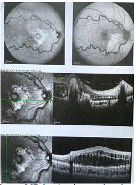
At examination, the visual acuity was counting fingers at 2 meters in the right eye and 10/10 in the left one, with no significant refractive error. Anterior segment examination was unremarkable and the intraocular pressure was 13 and 15mm Hg, respectively Ocular funduscopy of the right eye showed an orange elevetad tumor of 3 disc diameters with dilated feeding vessel and tortuous draining vein and significant macular edema with lipid exudates (Figure1)

Figure 1: Right eye fundus: nette résorption des exsudats maculaires , l’aspect des vaisseaux est moins tortueux apres un an.
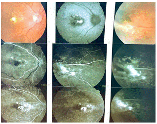
Fluorescein angiography revealed hyperfluorescence of the tumor in the early phase, whereas marked dye leakage was noted in a late phase. The Optical Coherence Tomography (OCT) confirmed the macular oedema (Figure 2). The diagnosis of retinal hemangioblastoma was established.

Figure 2: Right eye OCT showing the macular edema.
The Brain MRI and the abdominal CT scan results were unremarkable. The genetic testing was in favor of VHL disease.
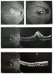
The patient had to undergo an argon laser photocoagulation of the retinal lesion. The laser burns were applied directly on the retinal hemangioblastoma one year after the trealment, the visual acuity in the rigjt eye was 2/10. The optical coherence tomography showed a large centrofoveolar exsudate and the fluorescein angiography revealed the regress of the retinal hemangioblastoma (Figure 3 and 4).

Figure 3: Right eye OCT: Evolution of the macular edema after trealment.

Figure 4: Retinal angiography showing the results 8 months after photocoagulation.
Discussion
Retinal hemangioblastoma is one of the most frequent tumors occurring during the course of VHL disease and can be responsible for significant visual impairment.
The disease results from germline mutations in the VHL tumor suppressor gene located on chromosome 3p25-p26, and the tumor development results from somatic inactivation of the remaining wild-type VHL allele [1].
The contribution of the ophthalmological examination in VHL is considerable, because the first manifestation of VHL is ophthalmic in 50% of patients.
On fundus evaluation, the lesions have an easily recognizable globular reddish appearance with a dilated feeding artery and a tortuous draining vein. The size of the retinal hemangioblastoma may be variable, ranging from a pinpoint lesion or a discrete vascular tuft to a large globular lesion [1].
The superior temporal quadrant is the predilection site for retinal hemangioblastoma [2]. The most common causes of vision loss are intraretinal exudation, exudative retinal detachment, hemorrhage, and epiretinal fibrosis [2].
Fluorescein angiography remains the gold standard for identifying small angiomas, juxta papillary angiomas or angiomas obscured by epiretinal membrane [3] Fluorescein angiography shows early hyperfluorescence and late leakage.
Successful treatment of peripheral tumors has been reported with laser photocoagulation, especially in smaller tumors, and with cryotherapy, brachytherapy, pars plana vitrectomy and diathermy [4].
Laser photocoagulation, brachytherapy and transpupillary thermoplasty have been shown to be effective in the treatment of optic disc hemangiomas, but all these approaches are risky and can result in permanent scotomas and poor clinical outcome due to the posterior location of the tumor and its proximity to the optic nerve [5].
Cryotherapy may be useful for larger peripheral lesions or associated exudative retinal detachment [12]. Brachytherapy reserved for larger lesions [6-8].
Retinovitreal surgery is to be considered for severe cases of retinal capillary hemangioblastoma associating serous RD, vascularized or non-vascularized pre-retinal fibrosis and neo-vessels on the tumor or pre-papillary [8,9].
In spite of its benign nature and classic slow-growing course, ocular hemangioblastoma may cause sight-threatening complications and remains a major cause of visual morbidity or sometimes blindness for patients with VHL disease. Early detection and treatment can change the visual prognosis [1].
According to french guidelines, yearly ophthalmic examination is indicated for patients with VHL disease under the age of 5 years. Between the age of 15 and 30 years, a funduscopy is recommended twice a year. After 30 years of age, a yearly examination is recommended [1].
Conclusion
Diagnostic delay due to the variability of clinical manifestations is one of the main difficulties encountered in the management of patients with VHL disease. The early diagnosis allows monitoring of lesions that can threaten the vital or visual outcome.
The ophthalmologist therefore has a major role to play in screening since 2/3 of patients with the disease present with retinal hemangioblastoma.
Author Statements
Conflict of Interest
The authors declare no conflict of interest.
Contribution of the Authors
All the authors participated in the care of the patient and the writing of the manuscript. All authors have read and approved the final version of the manuscript.
References
- Dollfus H, Massin P, Taupin P, Nemeth C, Amara S, et al. Retinal hemangioblastoma in von Hippel-Lindau disease: a clinical and molecular study. Invest Ophthalmol Vis Sci. 2002; 43: 3067-74.
- Webster AR, Maher ER, Moore AT. Clinical characteristics of ocular angiomatosis in von Hippel-Lindau disease and correlation with germline mutation. Arch Ophthalmol. 1999; 117: 371-8.
- Nabih O, Hamdani H, El Maaloum L, Allali B, El Kettani A. Retinal angioma of Von hippel-lindau disease: A case report. Ann Med Surg (Lond). 2022; 74: 103292.
- McCabe CM, Flynn HW Jr, Shields CL, Shields JA, Regillo CD, et al. Juxtapapillary capillary hemangiomas. Clinical features and visual acuity outcomes. Ophthalmology. 2000; 107: 2240-8.
- Saitta A, Nicolai M, Giovannini A, Mariotti C. Juxtapapillary retinal capillary hemangioma: new therapeutic strategies. Med Hypothesis Discov Innov Ophthalmol. 2014; 3: 71-5.
- Atebara NH. Retinal capillary hemangioma treated with verteporfin photodynamic therapy. Am J Ophthalmol. 2002; 134: 788-90.
- Chan CC, Collins AB, Chew EY. Molecular pathology of eyes with von Hippel-Lindau (VHL) Disease: a review. Retina. 2007; 27: 1-7.
- Kreusel KM, Bornfeld N, Lommatzsch A, Wessing A, Foerster MH. Ruthenium- 106 brachytherapy for peripheral retinal capillary hemangioma. Ophthal mology. 1998; 105: 1386-92.
- Mennel S, Meyer CH, Callizo J. Combined intravitreal antivascular endothelial growth factor (Avastin) and photodynamic therapy to treat retinal juxtapapillary capillary haemangioma. Acta Ophthalmol. 2010; 88: 610-3.
- Gaudric A, Korobelnik JF, Quentel G, Coscas G. Traitement du décollement de rétine au cours de l’angiomatose de von Hippel. Bull Soc Ophtalmol Fr. 1987; 87: 1357-8.