Clinical Image
Austin J Clin Ophthalmol. 2023; 10(6): 1163.
Christmas tree Cataract
Najoua El Moubarik*; Zeinabou Hmeimett
HmeimettDepartment of Ophthalmology “A”, Ibn Sina University Hospital (Hôpital des Spécialités), Mohammed V University, Rabat, Morocco
*Corresponding author: Najoua El Moubarik Department of Ophthalmology “A”, Ibn Sina University Hospital (Hôpital des Spécialités), Mohammed V University, Rabat, Morocco. Email: najoua.elmoubarik@gmail.com
Received: August 24, 2023 Accepted: September 26, 2023 Published: October 03, 2023
Clinical Image
We report the case of a 68-year-old patient, with no particular pathological history, who consulted for a progressive decline in visual acuity over the past 4 years.

Figure 1: Overall survival, autologous stem cell transplant (ASCT) versus no ASCT (p=0.12).
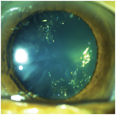
Ophthalmological examination of the right eye showed, after pupillary dilatation, multicolored scintillating opacities in the crystalline lens, appearing as colored lights decorating a christmas tree. The rest of the biomicroscopic examination was normal. The diagnosis of Christmas tree cataract was made. Christmas tree cataract is rare. This form of cataract is characterized by fine, polychromatic, reflective needle-point deposits in the deep cortex and nucleus. These deposits consist of cysteine, one of the least soluble amino acids. This cataract is often related to age and is said to be different from polychromatic cataract, associated with type 1 myotonic dystrophy, in which the particles are smaller in size.