Research Article
Austin J Clin Ophthalmol. 2024; 11(1): 1173.
Liberty® by Medicontur, a Complex in Structure, Hydrophilic Intraocular Lens in the Correction of Eyes with Preoperative Myopia
Adam Cywinski*; Paulina Bazgier
Silesian Eye Treatment Centre, Poland
*Corresponding author: Adam Cywinski Silesian Eye Treatment Centre, Okrezna 11, 44-240 Zory, Poland. Tel: 0048502137635 Email: adamcyw@gmail.com
Received: November 29, 2023 Accepted: January 02, 2024 Published: January 09, 2024
Abstract
A retrospective analysis of visual function in patients with preoperatively diagnosed myopia, after implantation of Liberty® intraocular lens, including the influence of some factors like a kind of surgery, values of angle alpha and kappa, pupil size, values of higher order aberrations generated by the lens on visual acuity to far and near distances was done. Lens removal and Liberty® implantation was done 47 eyes, including in 13 patients in both eyes. In 7 eyes, it was a toric lens. A mean preoperative value of myopia was -6.72 Dsph. Cataract removal accounted for 55% of all procedures performed, the rest being Refractive Lens Exchange. Preoperative pupil size was between 3.6 and 6,4 mm. The average postoperative visual acuity to far distances was 0.13 (logMar), and to near distances was 0.51 (Snellen). Rare cases of insufficient quality of vision to intermediate distances, when the postoperative pupil size was greater than 4 mm, resolved after increasing light intensity in the room. A bigger values of higher order aberrations corresponded with lower values of preoperative visual acuity to far distances. A bigger values of angle alpha corresponded with lower values of postoperative visual acuity to far distances, which were significantly higher in cataract group. Visual acuity to far distances was also better in eye with bigger pupil size. A specific structure of the lens allows patients to see good to the near distances, even if the light intensity not perfect.
Keywords: Angle kappa; Angle alfa; Cataract; Diffractive lens; EDOF; Higher order aberrations; Myopia; Refractive lens exchange
Abbreviations: EDOF: Extended Depth of Focus; VA: Visual Acuity; HOA: Higher Order Aberrations; RLE: Refractive Lens Exchange; LVC: Laser Vision Correction; UBVA: Uncorrected Best Visual Acuity; BCVA: Best Corrected Visual Acuity; LRI: Limbal Relaxing Incision
Introduction
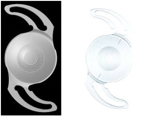
Liberty® intraocular lens is a product made by Medicontur Company. Its complex structure combines the features of a diffractive, trifocal lens with the EDOF structure (Figure 1a,b).

    Figure 1: An example of spheric model of Liberty® lens (left) 1b. An example of toric model of Liberty® lens (right). The flexible loops provide good stability into the capsula.

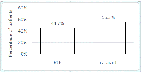
Figure 1: Graphical representation of Liberty lens implantation in two groups of patients (%).

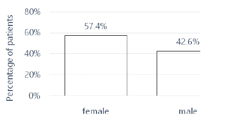
Figure 2: Graphical representation of Liberty lens implantation depending on gender.
The structure complexity only covers the central area, 3mm in diameter compared to 6mm, which is the total dimension of the lens optics. This structure allows us to obtain better quality of vision to far distances. The addition of +1.75 dioptres to intermediate distances and total addition +3.5 dioptres ensures good vision to near distances. As Fernandez points out, the lens allows you to read even in low light conditions, which constitutes its strong advantage. On the other hand, the author points out that in low light conditions, when the pupil size increases above 3.5 mm, visual acuity to intermediate distances may deteriorate. In order to obtain good quality of vision, light intensity should be increased, which automatically reduces the pupil size [1].
A large addition to near distances becomes important in the case of correction of aphakia in eyes that were preoperatively myopic. Patients with severe and moderate myopia, due to very good vision to near distances, not requiring additional correction, will pay particular attention to the quality of vision to near distances. The complex structure of the lens, using EDOF, has its limitations. They include a small pupil size, assessed under photopic conditions. Implantation of lenses with this structure in the case of a small pupil size significantly increases the risk of poor vision to far distances [2,3].
Taking into account the author’s experience, it is difficult to “satisfy” the patient in the field of vision to near distances when preoperative myopia exceeded -3,0 -4,0 dioptres. After obtaining emmetropia with the use of a complex intraocular lens with a large (3-4 dioptres) reading addition, patients almost always express their satisfaction with regaining vision to far distances, but often report that vision to near distances is worse than before the procedure. What is the cause of that and what influences it?
Myopic patients have to re-learn to read at a distance of about 40 cm, just like patients with emmetropia do. At the same time, they must “forget” that before the procedure, without the need for additional correction, they could read the text from very close distances, i.e. 5-10 cm. However, this takes time, often even several weeks.
Objective
Evaluation of the quality of vision and VA before and after the procedure, with particular emphasis on vision to near distances, in patients undergoing postoperative aphakia correction with the use of the Liberty® lens by Medicontur. Other objectives are to determine whether there are correlations between the obtained VA and the reason for the procedure and the parameters of the eye structure, taking into account the size of the preoperative refractive error, the values of angles kappa and alpha, the size of lens-generated HOA using iTrace analyzer and the pupil size.
Methods
Inclusion criteria: Preoperative, axial and mixed myopia, i.e. axial-refractive, also with concomitant corneal astigmatism. Two groups of patients were included: with diagnosed cataract and who wish to get rid of their refractive errors as part of RLE. High myopia was not a contraindication to the procedure. Confirmed loss of accommodation if eligible for RLE.
The qualifying examinations included the assessment of the condition of the cornea (including count of endothelial cells), the lens, the retina and the optic nerve. As a standard, the values of angles kappa and alpha, cornea-generated HOAs, values and intraocular pressure were assessed. Preserved, normal pupil function is an essential element of the inclusion criterion in the group.
Three patients (3 eyes) with diagnosed complicated cataract, who previously were operated on using posterior vitrectomy surgery because of diagnosed retinal detachment, and one patient (bilaterally) after of myopia LVC were also qualified for the procedure. The condition for qualifying patients after LVC was the presence of low cornea-generated HOAs.
A large preoperative pupil size, above 5.5 mm, examined in photopic conditions, was not a contraindication to the procedure due to the “favourable” structure of the Liberty lens.
The main reasons for consenting to the procedure by patients include the wish to improve vision in patients with diagnosed cataract and to get rid of refractive errors.
Exclusion Criteria
A small pupil size, less than 3.5 mm. Dysfunction of pupillary sphincter. Poor visual acuity caused by dysfunction to one of the eye structures, with poor prognosis as to obtaining good postoperative visual acuity. High cornea-generated HOA values.
All procedures were performed in a private medical centre - Silesian Eye Treatment Centre in Zory (Poland) by one surgeon. In each case the lens was calculated using optical biometry. Phacoemulsification (eyes with cataract) or phacoaspiration (RLE) procedures were performed with subsequent placement of the lens in the capsule. A toric lens was chosen for implantation in the case of corneal astigmatism with a value =1.0 Dcyl.
Visual acuity to far distances was assessed using EDTRS charts, and to near distances using Snellen charts.
Patient consent.
Each patient has written informed consent for inclusion of their clinical and imaging details in the manuscript for the purpose of publication.
The Ethics Committee (from Silesia) accepted both the implantation of this lens in human eyes due to its previous registration and the form of implantation.
The study was performed in accordance with the Helsinki Declaration of 1964, and its later amendments.
Data Availability Statement
All data, which were used to make statistics, can be available to verify the information presented in this article, but these data cannot be used in other studies without the express consent of the authors of this work. They are not publicly available, but it doesn’t mean all data used in this article are not true.
Statistical Tests Used
The significance level was p=0.05. Accordingly, results of p<0.05 will indicate significant correlations between the variables. Calculations were made in the statistical environment R ver.3.6.0, PSPP program and MS Office 2019.
Results
All procedures were performed between 2020 and 2023. The follow-up period is 6 to 30 months, 13 months on average. The values obtained during the last appointment, in a period of not less than 6 months, were used for the analysis of postoperative visual acuity. Information on the study group, including sex, percentage of cataract removal and RLE, type of lens implanted, and eye dominance are included in Table 1.
    group

    N

    %


    RLE

    21

    44.70%


    cataract

    26

    55.30%


    sex

    N

    %


    female

    27

    57.40%


    male

    20

    42.60%


    eye

    N

    %


    L

    23

    48.90%


    R

    24

    51.10%


    lens type

    N

    %


    Liberty

    40

    85.10%


    Liberty Toric

    7

    14.90%


    dominant eye

    N

    %


    D

    18

    56.30%


    N

    14

    43.80%


    type of defect

    N

    %


    myopia

    40

    85.10%


    myopic astigmatism

    7

    14.90%
Table 1: Group characteristics.
The percentage of both groups, i.e. cataract and RLE, is presented, below in Graph 1.

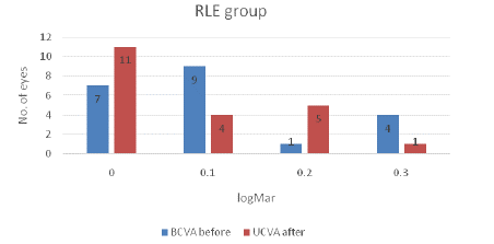
Figure 1: A comparison of visual acuity to far distances (logMar) examined before surgery with best correction to result gained after the surgery, but without correction, in RLE group.
The majority of the study group were women, as shown in Graph 2.

Graph 2: A comparison of visual acuity to far distances (logMar) examined before surgery with best correction to result gained after the surgery, but without correction, in cataract group.
Women more often decided to have the procedure performed due to the presence of a Refractive Error (RLE).
Below, in Table 2, for the study group of N=47 patients, descriptive statistics are presented, taking into account the mean, minimum and maximum values, as well as the values of median age and individual vision parameters before and after the procedure.
    Variable

    N

    M

    SD

    Min

    Max

    Me


    age [yrs]

    47

    49.53

    10.66

    27.00

    73.00

    49.00


    biometry [mm]

    47

    25.62

    1.34

    22.42

    27.92

    25.83


    spherical equivalent, preop. [D]

    46

    -6.72

    3.53

    -16.50

    -0.38

    -6.01


    pupil size, preop. [mm]

    45

    4.90

    0.72

    3.20

    6.40

    5.00


    UBVA to far, preop. [logMAR]

    27

    1.23

    0.48

    0.20

    1.80

    1.50


    BCVA to far, preop. [logMAR]

    45

    0.24

    0.21

    0.00

    0.70

    0.20


    UCBVA to near, preop. [Snellen]

    37

    0.50

    0.00

    0.50

    0.50

    0.50


    BCVA to near, preop. [Snellen]

    44

    0.50

    0.00

    0.50

    0.50

    0.50


    spherical equivalent, postop. [D]

    47

    -0.46

    0.48

    -2.00

    0.38

    -0.38


    pupil size, postop. [mm]

    47

    4.81

    0.61

    3.60

    6.30

    4.80


    UBVA to far, postop. [logMAR]

    46

    0.15

    0.13

    0.00

    0.40

    0.20


    BCVA to far, postop. [logMAR]

    47

    0.14

    0.13

    0.00

    0.50

    0.10


    UBVA to near, postop. [Snellen]

    46

    0.52

    0.06

    0.50

    0.75

    0.50


    BCVA to near, postop. [Snellen]

    47

    0.50

    0.01

    0.50

    0.55

    0.50


    kappa angle [mm]

    45

    0.31

    0.13

    0.02

    0.66

    0.33


    alpha angle [mm]

    45

    0.34

    0.13

    0.09

    0.65

    0.34


    HOA by lens [μm]

    47

    0.51

    0.55

    0.07

    3.38

    0.32


    HOA by cornea [μm]

    44

    0.21

    0.09

    0.06

    0.45

    0.18


    Note: N: Counts; M: Mean; SD: Standard    Deviation; Min: Minimum; Max: Maximum; Me: Median;    UBVA: Uncorrected Best Visual Acuity; BCVA: Best Corrected Visual Acuity; HOA:    High Order Aberrations
Table 2: Descriptives.
The parameters taken into account includes age, eyeball length, spherical equivalent value, pupil size, visual acuity to far and near distances, uncorrected and with best correction, tested before and after the procedure, as well as values of angles kappa and alpha, and cornea- and lens-generated HOAs. All the parameters are included in table 2.
The patients’ age was quite varied, ranging from 27 to 73. The eyeball length ranged from 22.42 mm to almost 28 mm, with the mean value of 25.6 mm, which does not fully coincide with the mean value of the spherical equivalent of the preoperative defect, being -6.72D (range -0.38D to -16.5D). Such a discrepancy may indicate the presence of a mixed refractive- axial error and/or corneal astigmatism.
Both kappa and alpha values were quite low, not exceeding 0.35mm, although there were values exceeding 0.65mm.
Visual acuity to far distances. Visual acuity and corneal astigmatism.
If UBVA values to far distances were worse in the postoperative period than BCVA values obtained before the procedure, attention was paid to the value of corneal astigmatism. The value =1.0 Dcyl resulted in the LRI procedure being performed in 5 eyes, which resulted in an improvement in visual acuity by an average of 1 row on the EDTRS charts and an improvement in the quality of vision.
BCVA values obtained before the procedure and UBVA after the procedure in the RLE group are presented in the form of Graph 3.
Although best correction of the existing error was applied in the preoperative period, in some cases visual acuity improved after the procedure. BCVA values obtained before the procedure and UBVA after the procedure in the cataract group are presented in the form of Graph 4.
For this group, it seems logical that visual acuity improved after the procedure. In 5 eyes, visual acuity was not = 0.4 logMar. These values were obtained in patients with, among others, progressive changes of retinitis pigmentosa (2 eyes), epiretinal membrane (1 eye), and diabetic changes, i.e. condition after posterior vitrectomy surgery (1 eye).
A detailed analysis of the obtained results in terms of visual acuity was performed later in the article.
Verification of statistical hypotheses
Hypothesis 1: The type of the procedure significantly affects the difference between pre- and postoperative visual acuity to far distances.
To test the assumed hypothesis, a two-factor ANOVA variance analysis with repeated measures was used (table 3).
    Effect

    SS

    df

    F

    p

    η²p


    Within Subjects Effects

    visual acuity

    0.18

    1

    11.72

    0.001

    **

    0.21


    visual acuity  group

    0.18

    1

    11.72

    0.001

    **

    0.21


    Residual

    0.66

    43


    Between Subject Effects

    group

    0.54

    1

    18.66

    <0.001

    ***

    0.30


    Residual

    1.24

    43


    Note: SS: Sum of Squares; df: Degrees of Freedom; F: Variance analysis test statistics; p: Statistical    significance; η²p: Partial Eta Squared;


      *p<0.05; **p<0.01; ***p<0.001
Table 3: The influence of the type of the procedure on the postoperative change in visual acuity to far distances - analysis of variance.
The study showed a statistically significant (p<0.05) simple effect in the form of a significant difference in visual acuity to far distances between examinations before and after the procedure. The interaction effect of the type of the procedure (visual acuity * group), i.e. the effect of the type of the procedure on the change in visual acuity, was also significant (p<0.05). In order to examine the differences in detail, Tukey post hoc test was carried out - a pairwise comparison (Table 4).
    Comparison


    visual    acuity

    group


    visual    acuity

    group

    MD

    SE

    df

    t

    p


    BCVA to far, preop.

    RLE

    -

    BCVA to far, preop.

    cataract

    -0.24

    0.05

    43.00

    -4.87

    < 0.001

    ***


    BCVA to far, preop.

    RLE

    -

    UBVA to far, postop.

    RLE

    -0.00

    0.04

    43.00

    -0.00

    1.000


    BCVA to far, preop.

    RLE

    -

    UBVA to far, postop.

    cataract

    -0.07

    0.04

    43.00

    -1.46

    0.470


    BCVA to far, preop.

    cataract

    -

    UBVA to far, postop.

    RLE

    0.24

    0.04

    43.00

    5.56

    < 0.001

    ***


    BCVA to far, preop.

    cataract

    -

    UBVA to far, postop.

    cataract

    0.18

    0.04

    43.00

    5.01

    < 0.001

    ***


    UBVA to far, postop.

    RLE

    -

    UBVA to far, postop.

    cataract

    -0.07

    0.04

    43.00

    -1.74

    0.317


    Note: MD: Mean Difference; SE: Standard Error; df: Degrees of Freedom; t: Test statistics; p: Statistical significance;


      *p<0.05; **p<0.01; ***p<0.001
Table 4: The influence of the type of the procedure on the postoperative change in visual acuity to far distances – a pairwise comparison.
As it results from the pairwise comparison, before the procedure, visual acuity to far distances was significantly different between patients with cataract and patients who underwent refractive lens exchange, while after the procedure the results were similar. In addition, patients undergoing refractive lens exchange did not report any improvement in visual acuity after the procedure, and among patients with cataract this change was statistically significant. Table 5, updated below, and presents the marginal means of individual measurements of visual acuity to far distances depending on the type of the procedure.
    95%    Confidence Interval


    visual    acuity

    group

    M

    SE

    Lower    limit

    Upper    limit


    BCVA to far, preop.

    RLE

    0.11

    0.04

    0.04

    0.18


    BCVA to far, preop.

    cataract

    0.35

    0.03

    0.28

    0.42


    UBVA to far, postop.

    RLE

    0.11

    0.03

    0.05

    0.16


    UBVA to far, postop.

    cataract

    0.18

    0.03

    0.12

    0.23


    Note: M: Mean; SE: Standard Error
Table 5: The influence of the type of the procedure on the postoperative change in visual acuity to far distances - marginal means.
The study showed that in the first examination, before the procedure, visual acuity to far distances among patients undergoing RLE was M=0.11 (SE=0.04), and among patients with cataract it was significantly worse and amounted to M=0.35 (SE=0.03). In the second examination, after the procedure, visual acuity in the RLE group did not change and was M=0.11 (SE=0.03), and in the cataract group it was M=0.18 (SE=0.03), and here the difference was not statistically significant (p >0.05). Thus, it was shown that visual acuity to far distance significantly improved only among patients with cataract.
The hypothesis was accepted - the type of the procedure significantly affects the change in visual acuity to far distances. Graphically, the result is presented below in the form of Graph 5.
Hypothesis 2: The type of the procedure significantly affects the difference between pre- and postoperative visual acuity to near distances.
To test the hypothesis, a two-factor ANOVA variance analysis with repeated measures was used to compare mean values of visual acuity to near distances (BCVA before the procedure and UCVA after the procedure) between examinations, taking into account the between-subject effect of the type of the procedure (Table 6).
    95%    Confidence Interval


    visual    acuity

    group

    M

    SE

    Lower    limit

    Upper    limit


    BCVA to near, preop.

    RLE

    0.50

    0.00

    0.50

    0.50


    BCVA to near, preop.

    cataract

    0.51

    0.01

    0.48

    0.54


    UBVA to near, postop.

    RLE

    0.50

    0.00

    0.50

    0.50


    UBVA to near, postop.

    cataract

    0.52

    0.01

    0.49

    0.55


    Note: M: Mean; SE: Standard Error
Table 6: The influence of the type of the procedure on the postoperative change in visual acuity to near distances - marginal means.
Before the procedure, visual acuity to near distances of patients undergoing RLE was M=0.50 (SE=0.00) and M=0.51 (SE=0.01) among those with cataract. In the second examination, after the procedure, visual acuity in the RLE group did not change and was M=0.50 (SE=0.00), and in the cataract group it was M=0.52 (SE=0.01); therefore, the differences were not statistically significant (p>0.05). Visual acuity did not change in a statistically significant way in any of the study groups.
The hypothesis was rejected - the type of the procedure does not significantly affect the change in visual acuity to near distances.
Hypothesis 3: There is a significant correlation between the preoperative pupil size, cornea- and lens-generated HOA values, angles kappa and alpha, and postoperative visual acuity to far and near distances uncorrected and best correct.
The analysed variables were quantitative variables, therefore the correlation coefficient was used. The type of coefficient used was determined by the nature of the distribution of variables, which was verified with the Shapiro Wilk test. Since all variables describing visual acuity were statistically significantly different from normal distribution, Spearman’s rank correlation coefficient was used (Table 7).
    UBVA to far,    postop. [logMAR]

    BCVA to far,    postop. [logMAR]

    UBVA to near,    postop. [Snellen]

    BCVA to near,    postop. [Snellen]


    pupil size, preop.    [mm]

    rho

    -0.215

    -0.381*

    0.124

    -0.058


    p

    0.155

    0.01

    0.421

    0.704


    HOA by lens [μm]

    rho

    0.231

    0.385**

    0.056

    0.005


    p

    0.122

    0.008

    0.71

    0.971


    HOA by cornea [μm]

    rho

    0.043

    0.028

    0.009

    -0.054


    p

    0.782

    0.855

    0.955

    0.728


    kappa angle[mm]

    rho

    0.01

    -0.065

    -0.192

    0.186


    p

    0.947

    0.673

    0.212

    0.222


    alpha angle [mm]

    rho

    0.321*

    0.376*

    0.117

    -0.07


    p

    0.032

    0.011

    0.449

    0.649


    Note: rho: Spearman correlation    coefficient; p: statistical significance,


      *p<0,05; **p<0,01; ***p<0,001
Table 7: Correlation between preoperative pupil size, HOA values, angles kappa and alpha, and visual acuity.
There was a statistically significant correlation (p<0.05) between postoperative visual acuity to far distances without correction and angle alpha. The correlation was moderately strong, as evidenced by the value of the rho coefficient <= 0.5. It was a positive correlation, which means that the larger the angle alpha, the worse the visual acuity (higher logMAR values).
There was also a statistically significant, moderately strong and negative correlation (p<0.05) between postoperative visual acuity to far distances with best correction and the preoperative pupil size. The larger the pupil size, the better the visual acuity.
There was also a statistically significant, moderately strong and positive correlation (p<0.05) between postoperative visual acuity to far distances with best correction, lens-generated HOA and angle alpha. The higher the lens-generated HOA value and the angle alpha, the worse the visual acuity. In the above scope, the hypothesis was accepted.
Hypothesis 4: There is a significant correlation between preoperative spherical equivalent and postoperative visual acuity too far and near distances uncorrected and with best correction.
There was no statistically significant correlation (p>0.05) between preoperative spherical equivalent and postoperative visual acuity too far and near distances without correction and with best correction. The hypothesis was rejected.
Hypothesis 5: There is a significant correlation between preoperative lens-generated HOA and preoperative uncorrected and best-corrected visual acuity to far distances in the refractive lens exchange group and in the cataract group.
The study used Pearson’s correlation coefficient and Spearman’s rank correlation coefficient.
In the group of patients undergoing RLE, no statistically significant correlation (p>0.05) was found between the lens-generated HOA value and preoperative visual acuity to far distances without correction and with correction.
In the cataract group, there was a significant correlation (p<0.05) between lens-generated HOA and preoperative visual acuity to far distances with best correction. The correlation was moderately strong, as evidenced by the value of the rho coefficient <= 0.5. It was a positive correlation, which means that the higher the lens-generated HOA value, the worse the preoperative visual acuity to far distances with correction. In this respect, the hypothesis was accepted.
Hypothesis 6: The type of the procedure significantly differentiates the preoperative lens-generated HOA value.
After verifying the assumptions of the normality of distribution, it was reasonable to use the non-parametric U-Mann- Whitney test, comparing the medians of the dependent variable in individual groups. The results of the Mann-Whitney U test for independent samples are presented in Table 8.
    Descriptives


    U

    p

    Min

    Max

    Me


    group

    HOA by lens    [μm]

    75

    <0.001


    RLE


    0.07

    0.54

    0.23


    cataract


    0.22

    3.38

    0.61


    Note: U: Test Statistics; p: Statistical Significance;  Me: Median; Min: Lowest Score; Max: Highest Score
Table 8: Differences in preoperative lens-generated HOA between groups selected by type of the procedure.
Patients with cataract were characterized by statistically significantly (p<0.05) higher preoperative lens-generated HOA value than patients who underwent refractive lens exchange. The distribution of variables is shown on the graph.
Hypothesis 7: There is a significant correlation between preoperative lens-generated HOA and the preoperative pupil size in the refractive lens exchange group and in the cataract group.
There was no statistically significant correlation (p>0.05) between the preoperative lens-generated HOA value and the preoperative pupil size in any of the study groups. The hypothesis was rejected.
Vision to intermediate distances
The assessment was performed in well-lit conditions and patients were asked to read a text from a computer with a standard font size at a distance of 80-100 cm. Apart from a few patients whose visual acuity to far distances was reduced, most of them had no problem reading the text.
Postoperative ailments
They were observed mainly in the first weeks after the procedure. The most common were: negative photopsia’s (4 patients), blurred vision looking at the computer in lower light conditions (3 patients), halo (2 patients), and blurred vision to near distances (5 patients). The latter ailment was not confirmed in the examination of visual acuity to near distances, assessed during follow-up appointments, and it subsided systematically over time due to the acceptance of vision while reading in the new conditions, i.e. at a distance of 35-40 cm.
Discussion
Taking into account the obtained results, the correction of postoperative aphakia with the use of the Liberty® IOL in eyes with preoperative myopia gives predictable, expected results. The improvement of postoperative visual acuity to far distances corresponding to the increase in the pupil size, while maintaining comparable pre- and postoperative visual acuity to near distances, gives the doctor and the patient what they expected, i.e. preservation of vision to near distances and regaining vision to far distances, without the need for correction. In the study group, the largest preoperative pupil size, assessed in photopic conditions, was 6.4 mm. With such a size, practically no multifocal, intraocular lens should be recommended due to the enormous risk of poor postoperative vision quality. Only monofocal, pure EDOF and Liberty® lenses can be used in such situations [3,4].
The beneficial effect of a large pupil size on vision to far distances give the doctor additional information about the possible impact of a small pupil size on achieving poorer postoperative vision to far distances.
….I can see far better than before the procedure… despite the lack of statistical significance of this information, some patients from the RLE group confirmed the improvement of vision to far distances after the procedure, which is shown in Graph 3.
The negative impact of large angle alpha on the quality of vision has already been described, therefore the assessment of this parameter in the preoperative qualification is extremely important. Large angle alpha in combination with a complex in structure intraocular lens will have a negative impact on vision to far distances and in scotopic conditions [5,6].
Another parameter that is not analysed in a standard way so far is the negative impact of high lens-generated HOA values on the decrease in visual acuity and the quality of vision to far distances. High lens-generated HOA values accompany cataract. High lens-generated HOA values are also the cause of reduced visual acuity and the quality of vision, mainly to far distances, which the author of this work described in two articles. The last one is titled “Congenital lens dysfunction as a new, undiagnosed cause of decreased visual acuity based on observation over a period of 3 years” [7,8].
Thinking about the correction of aphakia in preoperatively myopic patients, implantation of a lens with a large addition to near distances should always be taken into account, so that the patient is able not only to regain vision without correction to far distances, but also to maintain vision to near distances, at least from a distance of about 35-40 cm. The gold standard would be to maintain vision to far and near distances in both eyes. In view of the above, the recently recommended correction with the use of intraocular lenses with the addition of vision to near distances at the level of +1.5Dsp and/or the creation of the so- called monovision or micromonovision is only a substitute for the expected postoperative benefits.
When choosing an intraocular lens model for a patient, we should be guided by the principle of “keeping the good the patient has received from nature and adding something beneficial”. Is “taking away” the patient’s binocular vision, and additionally stereoscopic vision, which he/she obtained before the procedure to far and near distances by appropriate correction of myopia, a good choice?
Micromonovision does not provide comfortable vision to far or near distances, and in the era of a large selection of lenses with a large addition to near distances, it is this type of lenses that should be recommended for patients with myopia.
Despite a wide range of intraocular lenses, there are only a few that could be offered to patients aged 40-50, because such patients most often wish to get rid of their refractive errors. According to the author of this study, lenses made of a hydrophobic material, in the case of which glistening phenomen has been confirmed, even with good structure parameters, resulting in good vision, should not be recommended. It is difficult to predict when and to what extent the glistening process will proceed [9].
Contrary to hyperopic eyes, eyes with diagnosed myopia are characterized by the smallest values of angle alpha, which means that the quality of vision, even after implantation of a highly complex lens, should be good [10-13].
Conclusions
Taking into account the structure of the Liberty® lens, this product meets the requirements allowing it to be recommended to patients with preoperative myopia. However, before performing the procedure, the patients should be informed about possible postoperative problems, most often transient, mainly related to vision to intermediate distances, in the case of a large pupil. This greatly facilitates the postoperative dialogue.
The statistical analysis showed that, comparing preoperative BCVA to postoperative UCVA, visual acuity to far distances significantly improved only among patients with cataract. The type of the procedure did not significantly affect the change in visual acuity to near distances and no statistical differences were found between preoperative BCVA and postoperative UCVA to near distances. The larger the preoperative angle alpha, the worse the postoperative UCVA to far distances, the larger the pupil size, the better the UCVA to far distances. The latter result is very favourable for patients with a larger pupil before the procedure. The higher the preoperative HOA value generated by the lens (mainly opaque) and the angle alpha, the worse the BCVA to far distances. The higher the lens- generated HOA value among patients with cataract, the worse the preoperative visual acuity to far distances with correction. Patients with cataract were characterized by significantly higher preoperative lens-generated HOA value than patients who underwent refractive lens exchange. Finally, there was no significant correlation between preoperative lens HOA values and pupil size in any of the study groups.
Values of higher order aberrations generated by the lens, should be examined, in every case of poor visual acuity, where the reason of deterioration is unknown.
Author Statements
Conflict Interest
Adam Cywinski, Paulina Bazgier, declare that they have no competing interests.
References
- Joaquín Fernández MD. Liberty, the most balanced trifocal IOL. Medicontur advertising article.
- Paik DW, Park JS, Yang CM, Lim DH, Chung TY. Comparing the visual outcome, visual quality, and satisfaction among three types of multifocal intraocular lenses. Sci Rep. 2020; 10: 14832.
- Cywinski A. The influence of angles kappa and alpha and pupil size on vision after implantation of Soleko evolve and lucidis lenses with a ”pure” EDOF structure. J Ophthalmol Adv Res. 2021; 2.
- Gundersen KG, Potvin R. Refractive and visual outcomes after implantation of a secondary sulcus intraocular lens with an extended depth of focus. Clin Ophthalmol. 2022; 16: 1861-9.
- Qin M, Ji M, Zhou T, Yuan Y, Luo J, Li P, et al. Influence of angle alpha on visual quality after implantation of extended depth of focus intraocular lenses. BMC Ophthalmol. 2022; 22: 82.
- Cervantes-Coste G, Tapia A, Corredor-Ortega C, Osorio M, Valdez R, Massaro M, et al. The influence of angle alpha, angle kappa, and optical aberrations on visual outcomes after the implantation of a high-addition trifocal IOL. J Clin Med. 2022; 11: 896.
- Cywinski A, Michulec M, Bloch D, Lubczyk A. Congenital lens dysfunction as a new, undiagnosed cause of decreased visual acuity based on observation over a period of 3 years. JOJ Ophthalmol. 2023; 10: 555777.
- Cywinski A. Usefulness of Itrace in diagnosing unclear cases of the deterioration in visual acuity. Congenital lens dysfunction as a new disease entity. Preliminary reports. J Biosci Biomed Eng. 2020; 1: 12.
- Henriksen BS, Kinard K, Olson RJ. Effect of intraocular lens glistening size on visual quality. J Cataract Refract Surg. 2015; 41: 1190-8.
- Velasco-Barona CMD, Corredor-Ortega CMD, Avendaño-Domínguez AMD, Cervantes-Coste GMD, Cantú-Treviño MP, Gonzalez-Salinas RMD. Impact of correlation of angle a with ocular biometry variables. J Cataract Refract Surg. 2021; 47: 1279-84.
- Baenninger PB, Rinert J, Bachmann LM, Iselin KC, Sanak F, Pfaeffli O, et al. Distribution of preoperative angle alpha and angle kappa values in patients undergoing multifocal refractive lens surgery based on a positive contact lens test. Graefes Arch Clin Exp Ophthalmol. 2022; 260: 621-8.
- Cywinski A. The influence of angles kappa and alpha and pupil size on vision after implantation of Soleko evolve and lucidis lenses with a ”pure” EDOF structure. J Ophthalmol Adv Res. 2021; 2.
- Cywinski A, Szyja O. Katy kappa i alfa w diagnostyce przedoperacyjnej pacjentów nadwzrocznych kwalifikowanych do wszczepienia soczewki wieloogniskowej z uzyciem technologii iTrace. OphthaTherapy. Ther Ophthalmol. 2022; 9: 90-7.