Case Report
Austin J Clin Ophthalmol. 2024; 11(1): 1175.
Orbital Cystic Lymphangioma in Children: A Case Report
Oumaima Boukhlouf¹*; Alain François Habimana¹; Taha Baiz²; Malik Boulaadas¹
¹Department of Maxillo-Facial Surgery, Ibn Sina Hospital (Hôpital des spécialités), Mohamed V University, Rabat, Morocco
²Department of Ophtalmology, Ibn Sina Hospital (Hôpital des spécialités), Mohamed V University, Rabat, Morocco
*Corresponding author: Oumaima Booukhlouf Department of Maxillo-Facial Surgery, Ibn Sina Hospital (Hôpital des spécialités), Mohamed V University, Rabat, Morocco. Email: boukhloufoumaima@gmail.com
Received: December 26, 2023 Accepted: January 25, 2024 Published: February 01, 2024
Abstract
Orbital lymphangioma is a rare benign tumor that represents 1-2% of all orbital orbital tumors [1]. These lesions are extremely serious not only because of its functional and aesthetic repercussions, but because it’s difficult to have a complete surgery due to the relationships [2].
The diagnosis is most frequently radiological and the management is often highly complex; involving both surgical and medical treatment.
Recurrence is frequent, even after successful surgical treatment.
In this work, we report the case of an orbital lymphangioma in a 6-month-old child with a good evolution after complete excision of the lesion.
Introduction
Lymphangiomas are benign congenital lesions which mostly affect the head and neck region [3]. They are progressive lesions, presenting as thin-walled and dilated vessels isolated from the arteriovenous circulation [2,4]. They are often diagnosed in early childhood, but may go undetected for several years. In other cases they can appear after a triggering factor such as trauma, upper airway infection [3,5,6].
Case Report
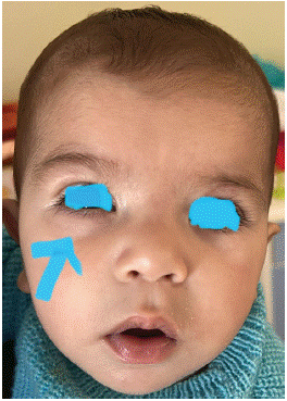
C.W, a 6-month-old boy with no previous pathological history, was brought to the emergency department of ophthalmology and maxillofacial surgery for lower palpebral swelling of the right eye associated with chronic tearing and recurrent conjunctivitis since birth. Ophthalmological examination revealed normal visual behavior, a glare reflex with positive photomotor reflex,good eye tone, preserved ocular motility and unilateral convergent strabismus of the right eye (Figure 1).

Figure 1: Lower eyelid swelling of the right eye.
Maxillofacial examination revealed a painless inferomedial palpebro-orbital tumefaction on the right, with no evidence of exophthalmos, proptosis or lateral dystopia. Ophthalmological examination of the left eye, ENT and general examination were normal. The Valsalva maneuver was negative.
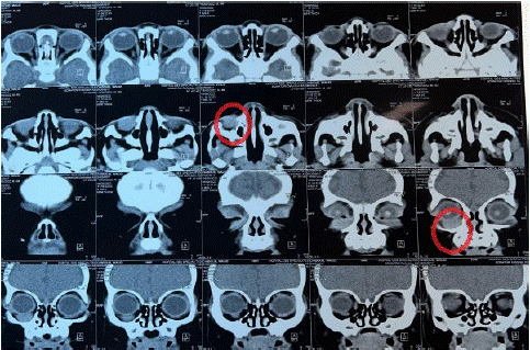
A cranio-orbital CT scan showed an extra-conic, homogeneous, well-limited intra-orbital mass, isodense poorly enhanced after contrast injection; measuring 10*16*14 mm. That mass had an intimate contact with the eyeball and the inferior rectus muscle and respecting the bone. (Figure 2).

Figure 2: Axial and coronal sections revealing an extraconal intraorbital mass in intimate contact with the eyeball, displacing the inferior rectus muscle, and respecting the adjacent bone.
The patient underwent laborious surgical removal of the tumor under general anesthesia, using a subciliary approach that respected the eyeball, its musculature and the orbital fat.
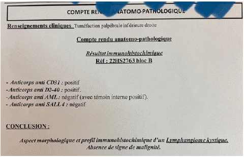
Anatomopathological analysis of the surgical specimen confirmed the diagnosis of lymphangioma cyst (Figure 3).

Figure 3: Anatomopathological analysis of the surgical lesion.
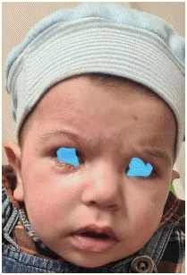
The postoperative course was straightforward, with good aesthetic and functional results (Figure 4).

Figure 4: Post-operative results on day 5 before suture removal.
At one year's follow-up, the patient hasn’t presented any tumor recurrence or ocular functional ocular disorders.
Discussion
Lymphangiomas are localized malformations of the vascular and lymphatic system that occur most frequently in the head and neck regions of children [7].
Their orbital-palpebral localization remains relatively rare, accounting for 1 to 2% of all orbital tumors [1].
They generally occur in the first decade of life, but may present late in the course of trauma to the orbit, recent infection of the upper respiratory tract, or hemorrhage of the eye [2,5,6].
In the orbit, lymphangiomas can be superficial (conjunctiva, eyelid), deep (intra-orbital) or mixed [2].
The tumor growth is slow and may remain silent for a long time [2]. The main symptom is exophthalmos, isolated or associated with other orbital signs such as ptosis, edema, asymmetry, proptosis, limitation of eye movements, decreased or lost visual acuity, amblyopia in a child [7].
However, intrinsic hemorrhage may occur within the tumor which increases its size, consequently leading to acute exophthalmos with occasionally the optic nerve compression, with or without diplopia and rapidly compromising ocular prognosis [2].
Lymphangiomas can also rapidly increase in size during episodes of respiratory, ENT infections, or superficial infections of the face; probably due to hyperplasia of the lymphocytic components [5,6].
Unlike hemangiomas, orbital lymphangiomas are not encapsulated and don’t respect precise anatomical boundaries [2]. They are sometimes associated with other angiomas, vascular anomalies or even other inflammatory diseases such as Inflammatory Bowel Disease [12].
Imagery has undeniable help in confirming the diagnosis [2].
Ultrasound associated with echo doppler may be useful, but magnetic resonance imaging remains the key exam in the diagnosis of orbital lymphangiomas. It determines the location, the appearance, the size and the relationship with orbital structures [3].
Orbital lymphangiomas are known for their difficult treatment, especially surgically; with a high recurrence rate. This is explained by the thinness of the cystic walls and their proximity to the eyeball and other orbital structures, which are all necessary for good vision, making the surgical procedure laborious [1,2,7].
The type of treatment also depends on lymphangioma size, cyst type and location [7]. Various procedures for cystic lymphangioma have been proposed in the literature but no consensus has yet been reached [8].
Some studies report regression of cystic lymphangiomas with different treatments: medical treatment with Sirolimus, surgery or sclerotherapy.
The indication of surgery is usually delayed until it becomes essential [8]. The surgery currently recommended is mainly conservative, and excision should be performed in the following situations: acute hemorrhage with optic nerve damage (as in our patient's case), significant exorbitism with corneal exposure or significant aesthetic disorders [2].
Drainage of the voluminous cysts, followed by filling of the cavity with tissue-glue to solidify the cysts and ensure a haemostatic effect may be proposed in certain cases, such as macrocysts [9,10].
Sclerotherapy consists in injection of sclerosing agent into the lesions aims to reduce lymphangioma size, asymmetry and pain, and to avoid surgical complications. The current products used are bleomycin /sodium tetradecyl sulfate (STS) / Picibanil (OK 432)/ ethanol [8].
Oral systemic medications such as aluminum hydroxide, bisphosphonates are also used to reduce size and symptoms in lesions that are difficult to access surgically or by injection [11].
Given its often locally and locoregionally infiltrative nature, sometimes with major and aesthetic repercussions, the prognosis for orbital lymphangiomas generally remains guarded [1].
Conclusion
Orbital lymphangiomas may be present from birth or appear in early childhood, which is why it is important to make this diagnosis in any child presenting with exophthalmos. Because of these sight-threatening complications, early and effective treatment is crucial to prevent functional and aesthetic prognosis.
References
- Derieux L, Catier A, Bertholom JL, Riffaud L, Charlin JF. 716 Prise en charge d’un lymphangiome kystique orbitaire chez une enfant de 3 ans. J Fr Ophtalmol. 2005; 28: 341-341.
- Moustaine MO, Hassoune M, Allali B, Elmaaloum L, Elkettani A, et al. Lymphangiome kystique intra-orbitaire de l’enfant: À propos d’un cas. J Fr Ophtalmol. 2019; 42: 411-4.
- Gmiha B, Boujemaa H, Haouari MA, Abdallah NB. Apport de l’IRM dans le diagnostic des lymphangiomes kystiques orbitaires. J Neurorad. 2017; 44: 106.
- Ducrey N. Les tumeurs vasculaires de l’orbite. Klin Monbl Augenheilkd. 1995; 206: 391-3.
- Jakobiec FA, Jones IS, editors. Vascular tumors, malformations and degenerations. Diseases of the orbit. Hagers-town. MD: Harper & Row. 1979: 229-308.
- Graeb DA, Rootman J, Robertson WD, Lapointe JS, Nugent RA, Hay EJ. Orbital lymphangiomas: clinical, radiologic, and pathologic characteristics. Radiology. 1990; 175: 417-21.
- Patel SR, Rosenberg JB, Barmettler A. Interventions pour le lymphangiome orbitaire. Cochrane. 2019.
- Montero, Beylerian M, et al. Lymphangiome Orbitaire Propos d’un cas Denis Congrès SFO. 2021.
- Patel KC, Kalantzis G, El-Hindy N, Chang BY. Sclerotherapy for orbital lymphangioma–case series and literature review. IV. 2017; 31: 263-6.
- Boulos PR, Harissi-Dagher M, Kavalec C, Hardy I, Codère F. Intralesional injection of tisseel fibrin glue for resection of lymphangiomas and other thin-walled orbital cysts. Ophthal Plast Reconstr Surg. 2005; 21: 171-6.
- Lerat J, Mounayer C, Orsel S, Bessede J, Aubry K. Lymphangiomes cervico-faciaux et prise en charge thérapeutique: étude clinique à propos de 23 cas. Ann Fr Oto-Rhino-Laryngol Pathol Cervico-Faciale. 2014; 131: A134.
- Sekhar LN, tariq F. Orbital lymphangiomas: surgical treatment and clinical outcome. World Neurosurg. 2014; 81: 710-1.