
Clinical Image
Austin J Clin Ophthalmol. 2024; 11(2): 1178.
Ocular Albinism: Smartphone Fundus Photography
Tanwar S¹*; Mishra S¹; Kumar B²
¹Department of Ophthalmology, King George Medical University, India
²Department of Dermatology, Rajiv Gandhi University of Health Sciences, India
*Corresponding author: Tanwar S Department of Ophthalmology, King George Medical University, Lucknow, India. Tel: 8950703083 Email: shashitanwar75.st@gamil.com
Received: January 12, 2024 Accepted: February 13, 2024 Published: February 22, 2024
Clinical Image
A 12 years old boy presented with diminution of vision, photophobia, and nystagmus in both eyes for the last 10 years. There was no history of ocular trauma or any systemic illness. Birth history was not relevant. There were no similar complaints in family. His best corrected visual acuity was 20/200 in both eyes. Slit lamp examination revealed iris hypopigmentation with transillumination defect. Fundus examination showed retinal depigmentation, conspicuously large Choroidal vessels, foveal hypoplasia, misrouting of the optic nerve fibers suggestive ocular albinism (Figure 1). Oculocutaneous Albinism (OCA) is a group of four autosomal recessive disorders [1]. In Ocular albinism; involvement is predominantly ocular, with normal skin and hair. Inheritance is usually X linked recessive. Reduction of melanin in the eyes results in reduced visual acuity caused by foveal hypoplasia and misrouting of the optic nerve fibers [2].
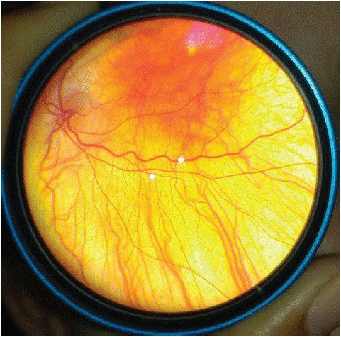
Figure 1: Smartphone fundus photograph of albinism showing retinal depigmentation, conspicuously large Choroidal vessels, foveal hypoplasia, misrouting of the optic nerve fibers.