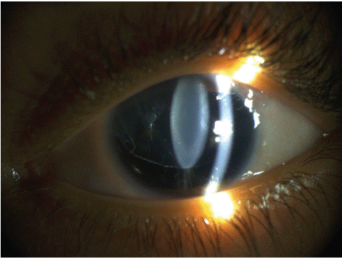
Clinical Image
Austin J Clin Ophthalmol. 2024; 11(2): 1179.
A Pediatric Presentation of Congenital Aniridia and Ectopic Lens
Meryem Sefrioui*; Hamza Lazaar; Hamidi Salma; Saad Benchekroun; Abdellah Amazouzi; Lalla Ouafa Cherkaoui
Department of Ophthalmology A, Rabat Specialty Hospital, University Mohamed V, Rabat, Morocco
*Corresponding author: Meryem Sefrioui Department of Ophthalmology A, Rabat Specialty Hospital, University Mohamed V, Rabat, Morocco. Tel: +212663058316 Email: meryemsefri@gmail.com
Received: January 23, 2024 Accepted: February 28, 2024 Published: March 06, 2024
Clinical Image
A 5-year-old, the youngest of three siblings, exhibited intense photophobia since birth. Under anesthesia, the left eye showed intraocular pressure of 21 mmHg, subtotal aniridia with persistent iridic remnants both inferiorly and superiorly, and an ectopic clear lens. The right eye had 25 mmHg intraocular pressure, subtotal aniridia with a residual iridic tissue inferiorly, and cataract. Fundoscopy revealed optic disc excavation and foveal reflex loss. Gonioscopy showed an open angle. No psychomotor delay or systemic anomalies were noted.
The patient underwent trabeculectomy for the left eye. Subsequently, the right eye underwent trabeculectomy combined with phacophagy, anterior vitrectomy, and intracapsular lens implantation.

Figure 1: Overall survival, autologous stem cell transplant (ASCT) versus no ASCT (p=0.12).
Aniridia is a rare condition characterized by complete or partial hypoplasia of the iris.
It can be isolated or associated with other ocular anomalies, most commonly congenital glaucoma (in over half of cases) and lens anomalies (cataract in 50-85% and ectopic lens in 50% of cases).