Letter to Editor
Austin J Clin Ophthalmol. 2024; 11(3): 1183.
Retinal Vein Thrombosis in Young Adult: Do not Omit a Primary Antiphospholipid Antibody Syndrome
Zaizaa M*; Maatalaoui F; Talamoussa B; El Bougrini Z; Bahadi N; Sahel N; Oumama J; El Kassimi I; Rkiouak A; Sekkach Y
Departement of Internal Medecine, Mohammed V Military Hospital, Rabat, Morocco
*Corresponding author: Zaizaa M Department of Internal Medecine, Military Hospital, Mohammed V University, Rabat, Morocco. Email: zaizaameryem@gmail.com
Received: February 13, 2024 Accepted: March 27, 2024 Published: April 03, 2024
Letter to Editor
Retinal vascular involvement is rare in AntiPhospholipid Antibody Syndrome (APS), and was first described in APS secondary to lupus disease. Retinal vascular damage can be a revelation of APS, especially in young patients. Its diagnosis is all the more important when the functional prognosis of the eye is at stake.
We illustrate the case of a young Moroccan patient who presented with vascular occlusion of the right eye during the course of primary antiphospholipid syndrome.
This 45-year-old man was admitted for a unilateral decrease of visual acuity in his right eye. His history included chronic smoking. He was not known to be diabetic or hypertensive, and had no complaints of recurrent bipolar aphthosis.
Three months prior to admission, he presented with a hazy sensation in the right eye. There was no raynaud's phenomenon or polyarthralgia, and the clinical examination was unremarkable, with no signs of connective tissue or inflammatory disease.
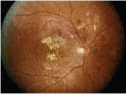
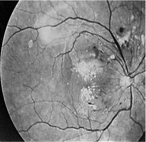
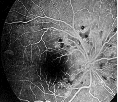
Fundus examination showed anterior uveitis with extensive vaso-occlusive lesions (Figure 1). Fluorescein angiography confirmed the diagnosis of retinal vein thrombosis in the right eye (Figure 2 & 3) brain imaging; echocardiography was normal.

Figure 1: Vaso-occlusive lesions.

Figure 2: Venous dilatation, arterial sheathing, retinal hemorrhages, cottony exudates: venous dilatation, arterial sheathing, retinal hemorrhages, cottony exudates

Figure 3: Severe retinal ischemia with perivascular dilation: severe retinal ischemia with perivascular dilation.
Biologically, there was no inflammatory syndrome (CRP 2.5 mg/L). The blood count was without abnormalities, as was the rest of the workup. The immunological workup was performed and showed negative antinuclear antibodies. The Elisa test for anti-β2-glucoprotin1 antibodies was positive at a high titre with two assays performed 12 weeks apart. The diagnosis of primary antiphospholipid syndrome was accepted and the patient received a treatment based on anticoagulant and the evolution was marked by a functional improvement one month later.
Antiphospholipid syndrome is an acquired thrombophilia characterized by the production of antoantibodies against a variety of phospholipids and phospholipid-binding proteins. These antibodies include anticardiolipin antibodies, b-2-glycoprotein-1 antibodies and lupus anticoagulan. When APS is diagnosed in the absence of any systemic disease that would account for elevation of these antibodies, the term primary antiphospholipid syndrome is used [1].
Retinal arterial or venous occlusion in primary or secondary APS is rare. Its frequency varies from 1% to 29% [2] depending on the study. Various ocular presentations have been reported in APS: central retinal vein or artery occlusion, diffuse retinal vaso-occlusive vasculopathy, sometimes severe [3].
Retinal vascular occlusions can be observed in a wide range of circumstances, including disseminated intravascular coagulation, diabetes, hereditary thrombophilias, churg-strauss syndrome, kawasaki disease and other conditions [2].
However, in some cases these occlusions are isolated and the etiology remains unknown, as in our patient cases’s. In such cases, antiphospholipid antibody syndrome should be considered, and a search made for antiphospholipid autoantibodies.
Retinal vein occlusion is a chronic disease with an unpredictable course. Complications include extensive ischemia and rupture of the hematoretinal barrier, leading to irreversible visual loss [3]; the presence of beta2GP1 antibodies, which were positive in our case, has been shown to be associated with thrombosis phenomena and are more specific markers than anticardiolipin antibodies in primary antiphospholipid antibody syndrome [2].
In the case of venous occlusion, the work-up is generally limited to a search for arterial hypertension, glaucoma or diabetes. A complete blood count and sedimentation rate are also performed to detect rare associated hemopathies or general inflammatory syndromes. There is no specific interest in performing an echodoppler examination of the neck vessels or an orbital CT scan [3].
Therapeutically, after a first spontaneous thrombotic episode, the high risk of thrombotic recurrence leads to the proposal of long-term anticoagulation [4]. our patient was put on HBPM with an early AVK relay and a target INR between 2-3.
young patients with unilateral retinal vein occlusion require regular monitoring, and should be made aware of the importance of therapeutic compliance and the need for emergency consultation in the event of a drop in visual acuity in the contralateral eye [5].
Retinal vein occlusion in APS is responsible for significant visual impairment, and its evolution is often unpredictable. Treatment remains controversial and generally disappointing. It is therefore important for ophthalmologists and internists to systematically test for antiphospholipid antibodies in young subjects presenting with retinal vascular occlusion.
References
- Ali O, Slagle WS, Hamp AM. Case report: Branch retinal vein occlusion and primary antiphospholipid syndrome. Optometry - Journal of the American Optometric Association. 2011; 82: 428–433.
- M Zitouni, F. Turki. Anticorps antiphospholipides et occlusion vasculaire rétinienne Annales de biologie clinique. 2002; 60: 278-91.
- Michel Paques, Alain Gaudric. Occlusions veineuses rétiniennes:Michel Paques, Alain Gaudric; Sang Thrombose Vaisseaux 2003; 15, n° 4: 215–9.
- Trojet S, Loukil I, El Afrit MA, Mazlout H, Bousema F, Rokbani L, et al. Bilateral retinal vascular occlusion during antiphospholipid antibody syndrome: a case report: J Fr Ophtalmol. 2005; 28: 503-507.
- Querques L, Terrada C, Souied E-H, Corrado A, Cantatore F-P, Querques G. Bilateral sequential central retinal vein occlusion associated with primary antiphospholipid syndrome. J Fr Ophtalmol. 2012; 35: 443.