Clinical Image
Austin J Clin Ophthalmol. 2024; 11(3): 1186.
Bilateral Peripapillary Intrachoroidal Cavitation
Sadiki*; Brarou H; Achegri Y; Elkhoyaali A; Fiqhi A; Mouzari Y; Oubaaz A
Department of Ophtalmology, Military Hospital Mohamed V Rabat, Morocco
*Corresponding author: Sadiki Department of Ophtalmology, Military Hospital Mohamed V Rabat, Morocco. Email: sadi.samah@gmail.com
Received: March 18, 2024 Accepted: April 19, 2024 Published: April 26, 2024
Clinical Image
Intrachoroidal cavitation is an abnormality of the choroid found most frequently in high myopic eyes. It was proposed that it’s the result of a choroid retraction away from the optic disc margin during staphyloma progression in myopic eyes. Peripapillary intrachoroidal cavitation are considered benign non progressive myopic lesions.

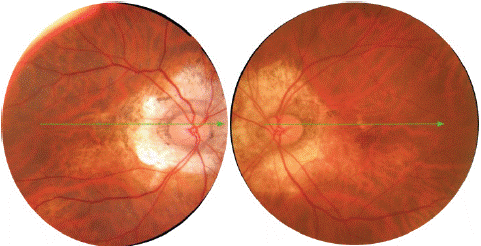
Colour fundus: Bilateral yellowish peripapillary lesion.

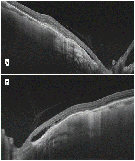
Colour fundus: Optical coherence tomography showing an hyporeflective space below the RPE in the right (A) and left (B) eye.
We report the case of a 65 years old male, who presented to the ophtalmological with a gradual decline in vision in both eyes. The ophtalmological examination found a best-corrected visual acuity (BCVA) was 2/10 in both eyes with the following refraction: -10,00 (-1,00 × 85°) in the right eye (OD) and -8.50 (-1,75 × 100°) in the left eye (OS). Slit lamp examination found a clear cornea, normal anterior chamber depth, intraocular pressure of 16 mmHg. Fundus exam showed a bilateral well-defined yellow lesion located surrounding the border of the disc. Spectral-domain Optical coherence tomography was performed and showed intrachoroidal hypo-reflective space.