Clinical Image
Austin J Clin Ophthalmol. 2024; 11(4): 1188.
Coralliform Cataract
Hazil Z*; Hasnaoui I; Boujaada A; Akannour Y; Serghini L; Hajji Z; Abdallah E
Department of Ophthalmology B, Rabat Specialty Hospital, CHU ibn Sina, Mohammed V Souissi University Rabat, Morocco
*Corresponding author: Zahira Hazil Department of Ophthalmology B, Rabat Specialty Hospital, CHU ibn Sina, Mohammed V Souissi University Rabat, Morocco. Email: zahira.hazil@gmail.com
Received: April 17, 2024 Accepted: May 09, 2024 Published: May 16, 2024
Clinical Image
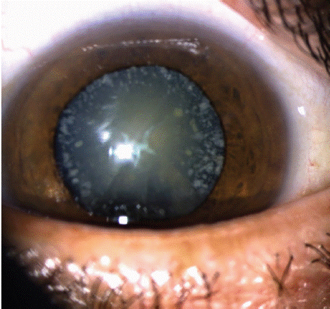
We report the case of a 50-year-old patient who consulted us for a bilateral decrease in visual acuity. On clinical examination, visual acuity was 2/10 in the right eye and finger movement in the left eye. Ocular tone was normal. Examination of the anterior segment revealed a cataract in the right eye, with whitish crystalline opacities predominating in the periphery, associated with a grade 2 nuclear cataract and sutural (Figure 1). Examination of the left eye revealed a subtotal cataract. The patient underwent phacoemulsification cataract surgery, which was uncomplicated, with simple postoperative follow-up. starting with the left eye and then the right.

Figure 1: Slit lamp image of a coralliform, nuclear and sutural cataract.
Coralliform cataract is a rare form of congenital cataract, characterized by bluish or whitish crystalline opacities, arranged in concentric layers with a radial center. It may be present at birth or develop in very early childhood, but is not diagnosed until adulthood. The condition is autosomal dominant, and may be caused by mutations in several genes (CRYBB2, CRYGD and MAF). Treatment is based on phacoemulsification of the lens, if visual acuity is impaired.
Author Statements
Conflicts of Interest
All authors declare that they have no conflicts of interest.