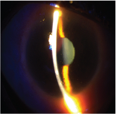
Clinical Image
Austin J Clin Ophthalmol. 2024; 11(4): 1189.
Anterior Uveitis Associated to Brimonidine: About A Case
Anass Boujaada*; Tlemcani Younes; Skalli Amine; Louai Serghini; Abdallah Elhassa
Department of Ophthalmology B, Hôpital des spécialités –IBN SINA Hospital University Center, Rabat, Morocco.
*Corresponding author: Anass Boujaada Department of Ophthalmology B, Hôpital des spécialités –IBN SINA Hospital University Center, Rabat, Morocco. Email: anass.boujaada@gmail.com
Received: April 25, 2024 Accepted: May 09, 2024 Published: May 16, 2024
Abstract
Brimonidine is commonly recognized as a culprit in ocular surface disease; however, its association with uveitis is less widely acknowledged. Here, we share significant clinical insights gleaned from one recent case of anterior uveitis linked to brimonidine.
Keywords: Brimonidine; Anterior uveitis
Introduction
Anterior uveitis, an inflammation of the iris and ciliary body of the eye, is an eye condition often associated with a variety of factors, including the use of certain topical medications. One of these substances, brimonidine, an agent commonly used in the treatment of glaucoma, has been linked to cases of anterior uveitis. This association between brimonidine and anterior uveitis raises significant clinical concerns, requiring a thorough understanding and appropriate management to prevent potential ocular complications in patients undergoing treatment with this drug.
Clinical Case
In January 2023, a 65-year-old woman, previously treated with topical brimonidine 0.2% in both eyes, presented with bilateral granulomatous anterior uveitis. She had no prior history of uveitis and was generally in good health. The patient was diagnosed with primary open-angle glaucoma in June 2021, and treatment commenced with levobunolol 0.5% twice daily. In October 2021, topical brimonidine 0.2% twice daily was added to both eyes. Fourteen months after initiating brimonidine, she experienced decreased visual acuity and was diagnosed with granulomatous anterior uveitis in both eyes, characterized by numerous "sheep fat" precipitates and anterior chamber tyndall. Levobunolol treatment continued in both eyes, while brimonidine drops were discontinued. The uveitis was managed with topical dexamethasone 0.1% every 2 hours for 4 days, followed by four times a day, and atropine 1% three times a day. After two weeks, inflammation resolved in both eyes, prompting cessation of atropine, and a gradual reduction in dexamethasone dosage over an additional two weeks.

Six weeks later, both eyes exhibited no signs of inflammation while solely on levobunolol. Ocular examination uncovered no underlying cause for the uveitis. Systemic assessments returned normal results, except for a C-reactive protein level of 15 mg/l, and she tested negative for HLA B27. Suspecting brimonidine as the culprit behind the uveitis, the right eye was reintroduced to topical brimonidine 0.2%. Within one week of resuming brimonidine, the right eye became uncomfortable, and after three weeks, fine granulomatous precipitates and discrete anterior chamber tyndall were observed. Brimonidine was promptly discontinued, and topical fluorometholone drops were administered three times daily for four weeks. Subsequently, both eyes have remained devoid of inflammation.
Discussion
The length of time between the start of brimonidine treatment and the onset of symptoms varied considerably from patient to patient. Some had been using brimonidine for just over a week, while others had been on it for up to four years before experiencing symptoms and receiving a diagnosis. This aligns with the timeframe indicated in a report by Beltz et al., which suggested a range of 1 week to 5 years for the onset of symptoms [1]. The extended period between the first use of brimonidine and the development of uveitis in many cases suggests that, in addition to a primary toxic reaction, there may also be involvement of a secondary cell-mediated immune response, with the drug possibly acting as an antigen or immune modulator [2]. It has been theorized that in patients undergoing long-term treatment, inflammatory cytokines within the vitreous humor might contribute to granulomatous uveitis [3]
Clinical signs of brimonidine-associated uveitis can vary widely, lacking a pathognomonic appearance akin to Fuch’s uveitis syndrome. The spectrum of clinical manifestations described previously includes simple conjunctival congestion, severe follicular conjunctivitis, blepharitis, punctate corneal epithelial erosions, anterior uveitis with corneal endothelial deposition (mutton fat keratic precipitates), significant anterior chamber activity (cells and flare), and posterior synechiae (PS). In our patient's case, there were no ps present, mirroring findings by Beltz et al. [1]. We primarily noted small, dispersed keratic precipitates, in contrast to the predominantly stellate keratic precipitates observed by Beltz et al. [1]. Our patient's age was 65 years, comparable to the mean age of 70 years (with a standard deviation of +/- 9 years) reported by Beltz et al. [1].
Conclusion
This form of anterior uveitis, while rare, holds significant clinical importance and should be kept in mind when managing glaucoma patients undergoing brimonidine treatment. Despite being treatable at its origin, challenges may persist, particularly concerning Intraocular Pressure (IOP) regulation. In some cases, achieving optimal IOP control may require glaucoma surgery even after the uveitis episode has been resolved.
References
- Beltz J, Zamir E. Brimonidine induced anterior uveitis. Ocul Immunol Inflamm. 2016; 24: 128–33.
- Moorthy RS, London NJS, Garg SJ, Cunningham ET. Drug-induced uveitis. Curr Opin Ophthalmol. 2013; 24: 589–97.
- Shin H-Y, Lee H-S, Lee YC, Kim S-Y. Effect of Brimonidine on the B cells, T cells, and cytokines of the ocular surface and aqueous humor in rat eyes. J Ocul Pharmacol Ther. 2015; 31: 623–6.