Case Report
Austin J Clin Ophthalmol. 2024; 11(4): 1191.
About a Case of Sutural Cataract
Anass Boujaada*; Younes Tlemçani; Lobna Robbana; Zahira Hazil; Louay Serghini; Abdallah Elhassan
Department of Ophthalmology “B”, Hôpital des Spécialités, IBN SINA Hospital University Center, Rabat, Morocco
*Corresponding author: Anass Boujaada Department of Ophthalmology “B”, Hôpital des Spécialités, IBN SINA Hospital University Center, Rabat, Morocco. Email: anass.boujaada@gmail.com
Received: April 25, 2024 Accepted: May 21, 2024 Published: May 28, 2024
Keywords: Cataract; Surgery; Syndromic cataract; Neuro-ophthalmology
Case Report
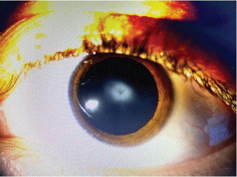
We report the case of a 13-year-old child, who consulted for a routine visual check. Visual acuity was 20/30 in both eyes. The biomicroscopic examination shows clear corneas, yet there were Y shaped lenticular opacities which followed the sutures of lens nucleus in both eyes. A neurological exam was required, which was normal with no signs of myotonia. Because the cataract did not cause a significant decrease in vision, it was not removed but monitored for progression.

Figure 1: Photograph of the anterior segment, showing thesutural cataract.
This form is diagnosed using the slit lamp, it does not have generally no functional impact. Sutural cataracts have a Y shape, which corresponds to the junction points of the lens fibers during embryogenesis: Y shape for the posterior suture and inverted Y shape for the anterior suture. it has an autosomal dominant inheritance.