Case Report
Austin J Clin Ophthalmol. 2024; 11(4): 1192.
Complete Paralysis of the Oculomotor Nerve III Caused by Ophthalmic Shingles
Tazi K*; Baiz T; Chefchaouni A; Elmoubarik N; Cherkaoui LO
Department of Ophthalmology A, Specialty Hospital of Rabat, Morocco
*Corresponding author: Kenza Tazi Department of Ophthalmology A, Specialty Hospital of Rabat, Morocco. Email: dr.ktazi@gmail.com
Received: April 26, 2024 Accepted: May 21, 2024 Published: May 28, 2024
Abstract
Ophthalmic herpes zoster, a manifestation of the reactivation of the varicella-zoster virus in the region of the trigeminal nerve, is primarily diagnosed based on the clinical characteristics of skin lesions and their localization. Ocular complications, including oculomotor paralysis, require early antiviral treatment to minimize ocular damage. We report the case of a 38-year-old woman presenting typical symptoms of ophthalmic herpes zoster, associated with complete oculomotor nerve III palsy and zoster encephalitis. Antiviral treatment with acyclovir led to a significant improvement. However, the patient retained corneal anesthesia resulting in the formation of a neurotrophic corneal ulcer. The risk of ocular complications associated with ophthalmic herpes zoster is real. The most serious are corneal complications and oculomotor paralysis, highlighting the crucial importance of early antiviral treatment to prevent these complications which can adversely affect the functional prognosis of the eye.
Keywords: Third nerve palsy; Ophthalmic zoster
Introduction
Ophthalmic shingles corresponds to the reactivation of the varicella-zoster virus (VZV) in the territory of the ophthalmic branch of the trigeminal nerve. The diagnosis is clinical based on the typical appearance of the skin lesions and their topography. Ocular damage can affect all structures of the eye including the oculomotor nerves, causing oculomotor paralysis which can lead to complete ophthalmoplegia. Antiviral treatment must be started early to limit ocular and neurological complications [1].
Clinical Case
We report the case of a 38-year-old diabetic patient on insulin therapy and without other medical, surgical or ophthalmological history, who consulted for a painful rash on the forehead and left hemiface eyelids associated with headaches.
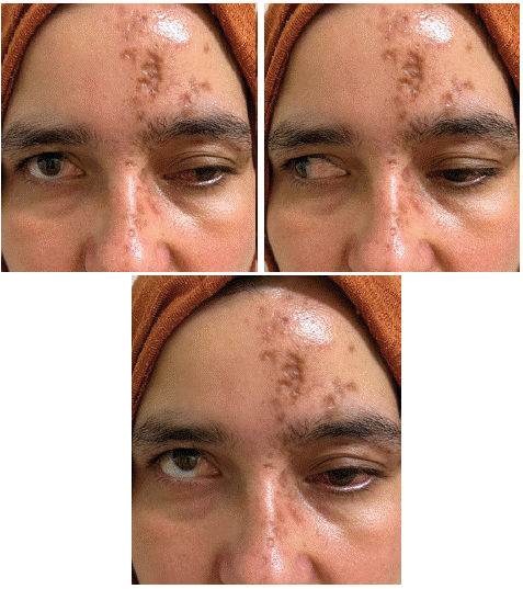
Examination of the left eye reveals ptosis associated with vesicular and crusty eyelid skin lesions extending over the entire territory of the V1 ophthalmic nerve. Palpation of the left hemiface reveals hypoesthesia on this same territory. The oculomotor examination reveals a complete extrinsic and intrinsic paralysis of the oculomotor nerve III, with light-reactive mydriasis associated with a limitation of adduction, elevation and lowering of the left eye. (Figure 1) The examination of the anterior segment and that of the fundus are unremarkable. Examination of the adelphic eye shows no abnormality and visual acuity is preserved in both eyes.

Figure 1: Crusted skin lesions of herpes zoster ophthalmicus with ptosis and limitation of adduction, elevation and lowering.
The patient's biological assessment revealed an inflammatory syndrome with positive IgG and IgM serology for Varicella-Zoster Virus (VZV). A lumbar puncture was performed, which revealed polynuclear neutrophilic pleocytosis with hyperproteinemia and hypoglycemia. A brain Magnetic Resonance Imaging (MRI) was performed, which returned without abnormality.
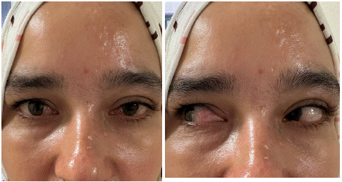
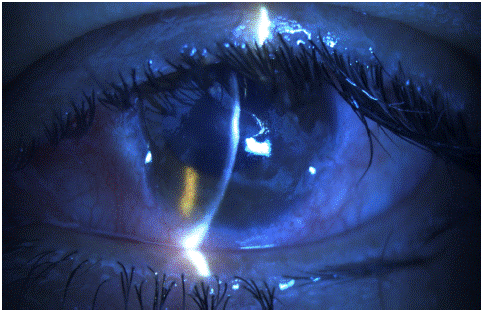
The diagnosis of ophthalmic zoster complicated by oculomotor paralysis and zoster meningoencephalitis was made. The patient received intravenous treatment with acyclovir. The evolution was marked by the progressive disappearance of ptosis, the almost complete recovery of the oculomotor paralysis and the healing of the skin lesions. (Figure 2) After three months, the patient presented with a painful red left eye with a drop in visual acuity, motivating her to consult. Clinical examination revealed diffuse conjunctival hyperemia with a central corneal ulcer and a total loss of corneal sensitivity in this eye. (Figure 3) Visual acuity in this eye was 4/10th. The diagnosis of neurotrophic ulcer secondary to ophthalmic shingles was made. The patient benefited from an amniotic membrane transplant combined with surface treatment with autologous serum eye drops. The evolution after 20 days was favorable with healing of the corneal ulcer and recovery of visual acuity in the left eye.

Figure 2: Regression of dermatological lesions and oculomotor paralysis after treatment with acyclovir.

Figure 3: Neurotrophic ulcer of the left eye secondary to ophthalmic shingles.
Discussion
Shingles ophthalmicus is an ocular infection induced by the reactivation of the varicella-zoster virus (VZV) which remains latent in the sensory ganglion of the trigeminal nerve following a primary viral infection with this same virus. The ophthalmic form represents 10 to 25% of all shingles [2]. The main risk factors are age over 60 and immunosuppression [3].
The diagnosis of ophthalmic zoster is clinical, based on the vesicular then crusty appearance of the rash, its painful nature and its characteristic topography located at the level of the ophthalmic territory of the trigeminal nerve (V1) and rarely exceeding the midline [4]. Certain additional examinations, in particular VZV serology, viral culture and detection of the virus by PCR, can be carried out in the event of diagnostic doubt regarding atypical forms [5].
In the absence of early antiviral treatment, ocular complications occur in 50% of cases and all structures can be affected [6]. The most common ocular manifestation during ophthalmic shingles is acute follicular conjunctivitis and the most serious is represented by corneal damage.
The latter can present in different aspects depending on the physiopathological mechanism and depending on the stage of the infection, which can go as far as functional loss of the eye. Approximately 25% of patients develop neurotrophic keratitis after shingles ophthalmicus. After-effect corneal anesthesia can lead to the appearance of chronic corneal ulcers, as illustrated by the case of our patient, who presented with a central neurotrophic ulcer three months after the episode of ophthalmic shingles [7]. Other ocular disorders may be associated with ophthalmic zoster, notably VZV anterior uveitis, VZV retinitis and chorioretinitis, or even ARN syndrome (accute retinal necrosis) and PORN syndrome (progressive outer retinal necrosis) [2].
Oculomotor paralysis during ophthalmic shingles is found in 5 to 31% of patients. Complete ophthalmoplegia is rare. The most common form is oculomotor nerve III paralysis. It can be partial or complete, both intrinsic and extrinsic, associating areflexic mydriasis, ptosis and limitation of adduction, elevation and lowering of the eye concerned. Cases of meningoencephalitis during shingles ophthalmicus have also been reported in the literature [8,9]. The case of our patient represents the typical form of complete paralysis of the oculomotor nerve III secondary to shingles ophthalmicus, associated with meningoencephalitis. -VZV encephalitis.
Treatment for shingles ophthalmicus should be started within the first 48 hours after the rash appears, to avoid the risk of eye complications. It is based on the systemic administration of antivirals. Several molecules have shown their effectiveness, in particular acyclovir, valaciclovir and famciclovir, the results of which have been demonstrated by several studies [10].
Conclusion
In conclusion, herpes zoster ophthalmicus, a manifestation of varicella-zoster virus reactivation in the trigeminal nerve region, presents significant diagnostic and therapeutic challenges. The clinical characteristics of skin lesions and their location play a crucial role in early diagnosis. Ocular complications, such as oculomotor paralysis and corneal damage, highlight the urgency of early antiviral treatment to prevent serious consequences on ocular function. The encouraging results of antiviral treatment with aciclovir, valaciclovir and famciclovir highlight the importance of their administration in the first 48 hours after the appearance of symptoms. Prompt multidisciplinary management is essential to optimize clinical outcomes and prevent potential complications associated with ophthalmic zoster.
References
- Yawn BP, Wollan PC, St Sauver JL, Butterfield LC. Herpes zoster eye complications: rates and trends. Mayo Clin Proc. 2013; 88: 562-70.
- Bourcier T, Borderie V, Laroche L. Zona ophtalmique. EMC - Ophtalmol. 2004; 1: 79-88.
- Tao BKL, Soor D, Micieli JA. Herpes zoster in neuro-ophthalmology: a practical approach. Eye Lond Engl. 2024.
- Pion B, Salu P. Herpes zoster ophthalmicus complicated by complete ophthalmoplegia and signs of pilocarpine hypersensitivity. A case report and literature review. Bull Soc Belge Ophtalmol. 2007: 23-6.
- Kalogeropoulos CD, Bassukas ID, Moschos MM, Tabbara KF. Eye and Periocular Skin Involvement in Herpes Zoster Infection. Med Hypothesis Discov Innov Ophthalmol J. 2015; 4: 142-56.
- Zografos L, Chamot L. [Ocular complications of zona ophthalmica]. Rev Med Suisse Romande. mars 1981; 101: 221-7.
- Harthan JS, Borgman CJ. Herpes zoster ophthalmicus-induced oculomotor nerve palsy. J Optom. 2013; 6: 60-5.
- Sanjay S, Chan EW, Gopal L, Hegde SR, Chang BCM. Complete unilateral ophthalmoplegia in herpes zoster ophthalmicus. J Neuro-Ophthalmol Off J North Am Neuro-Ophthalmol Soc. 2009; 29: 325-37.
- Tofade TO, Chwalisz BK. Neuro-ophthalmic complications of varicella-zoster virus. Curr Opin Ophthalmol. 2023; 34: 470-5.
- Cobo M. Reduction of the ocular complications of herpes zoster ophthalmicus by oral acyclovir. Am J Med. 1988; 85: 90-3.