
Case Report
Austin J Clin Ophthalmol. 2024; 11(5): 1193.
Topical Insulin Collyrium. Our Experience Treating Two Cases of Neurotrophic Ulcer with Anterior Chamber Fibrinous Reaction
Damian Garcia-Teillard, MD¹*; Salvador Garcia-Delpech, MD, PhD²; Sol Paula Cortes De Miguel, Pharm, MSc³; M Angeles Barranco-Romero, MD¹
¹Department of Ophthalmology, Punta Europa University Hospital, Algeciras, Spain
²AIKEN Ophthalmology Clinic, Valencia, Spain
³Department of Pharmacy, Punta Europa University Hospital, Algeciras, Spain
*Corresponding author: Damian Garcia-Teillard Department of Ophthalmology, Punta Europa University Hospital, Algeciras, Carretera de Getares S/N, Algeciras, 11207, Spain. Tel: +34619443173 Email: damiangteillard@gmail.com
Received: May 21, 2024 Accepted: June 10, 2024 Published: June 17, 2024
Abstract
Purpose: We report two cases of neurotrophic ulcers secondary to type II Diabetes involving an anterior chamber fibrinous reaction. Ulcers were treated using insulin collyrium 1 IU/mL.
Methods: Two diabetic patients with neurotrophic corneal ulcers (hyposensitivity confirmed with the air Cornea Esthesiometer Brill) showed anterior chamber fibrinous reaction and severe inflammatory signs. Insulin collyrium 1 IU/mL was administered in both cases to obtain a faster epithelization process, resolve the ulcer, and start anti-inflammatory therapy with topical dexamethasone. Oral and topical antibiotics were also administered as per our hospital’s protocol in patients showing corneal ulcers associated to fibrinous reaction in anterior chamber.
Results: Insulin collyrium helped in the epithelization and healing of the ulcers. One week later, we started treatment with topical dexamethasone phosphate collyrium twice a day, ceased the inflammatory process and promoted resorption of the fibrinous reaction in anterior chamber.
Conclusions: The use of topical insulin in treating corneal ulcers is still a relatively new and emerging treatment option. Up to date, the available published results of are promising and suggest that topical insulin may be a viable alternative or adjunctive treatment option for neurotrophic corneal ulcers. In these two cases, insulin collyrium helped in the epithelization process of the neurotrophic ulcers so that anti-inflammatory therapy with topical dexamethasone phosphate collyrium could be started as soon as possible and resolve the anterior chamber fibrinous collection
Keywords: Neurotrophic corneal ulcer; Insulin collyrium; Anterior chamber fibrine; Diabetic disease
Introduction
Corneal ulcers are a common and potentially serious eye condition that can cause pain, redness, blurred vision, and even vision loss if left untreated [1,2]. Treatment for corneal ulcers typically involves the use of antibiotics, antifungal or antiviral medications, and corticosteroids (although used sometimes and very carefully) to control the infection and promote healing [1,2]. However, recent research has shown that topical insulin may also be a promising treatment option for corneal ulcers [3,4].
Titone et al. Reported in 2018 [5] the homeostatic effect topical insulin could have on corneal epithelization, promoting PI3K-Akt pathway and stimulating IGF-1R and IR receptors in stromal and epithelial corneal cells. Topical insulin could reduce inflammation, and promote faster healing of corneal ulcers, which has implications for corneal ulcers.
David Diaz-Valle et al. reported in Acta Ophtalmologica [6] results from their clinical trial where they tested treatment for persistent epithelial defects with topical insulin 1IU/ml. They concluded on the effectivity oft this topical treatment in promoting healing of persistent epithelial defects.
In addition to topical insulin, other novel treatments for corneal ulcers are also being explored, such as stem cell therapy and gene therapy [4]. These new treatment options offer hope for patients with corneal ulcers who may not respond to traditional treatments or who may experience adverse side effects.
Case Presentation
In this work we present two clinical cases of neurotrophic ulcers with anterior chamber fibrinous reaction, treated with topical insulin collyrium. As in our hospital this treatment is “out of label”, both patients signed an informed consent to be treated with it.
Our first patient is a 70-year-old woman, suffering from type II Diabetes Mellitus and diagnosed with secondary chronic type III neurotrophic keratopathy, not feeling air pulse at any intensity (diagnosed with Corneal Esthesiometer Brill, Bill Engines Calle Munner 10, Barcelona 08022, SPAIN) in right eye. right eye had a previous history of three retinal detachments that had undergone multiple vitrectomy surgeries, with scleral buckle and silicon oil (Visual Acuity in previous reports 20/400 on Snellen chart) and glaucoma treated with topical prostaglandins (Bimatoprost 0.3 mg/mL twice a day). She visited our ophthalmology emergency unit reporting total vision loss and hiperoemia.
Upon admission, the patient’s visual acuity in the right eye was of “moving hands” at less than 50 cm and 20/50 (Snellen chart) in the left eye. Physical examination showed eyelid edema with severe inflammatory signs such as intense hyperemia, hypotony and fibrinous collection in the anterior chamber with hyphema (Figure 1a). The cornea Showed a central ulcer affecting more than 1/3 of its total area, fluorescein positive testing, and clearly infiltrated (Figure 1b). First patient presented corneal hyposthesia, not feeling any air impulse at pressure any pressure (CEB esthesiometer). Corneal ulcer samples (frotis) were taken for microbiological analysis.
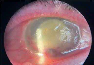
Figure 1a: Patient 1 upon admission. Intense hyperemia, hypotony and fibrinous collection in anterior chamber with hyphema.
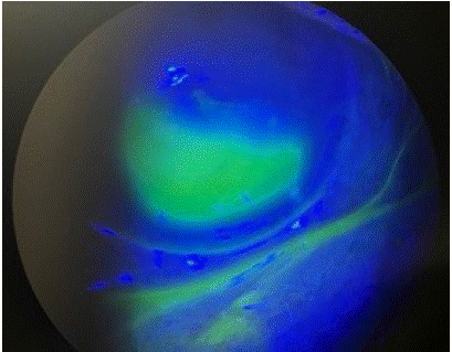
Figure 1b: Patient 1 upon admission. Corneal ulcer fluorescein positive testing.
Our patient did not report any infectious contact and was not a contact lens user. A differential diagnosis was made between infectious and inflammatory hypopyon. Bimatoprost was suspended and substituted with oral acetazolamide (250 mg once daily). We administered intravitreal antibiotics (Ceftazidime 2 mg/0.1mL and Vancomycin 1 mg/0.1mL) in addition to topical Vancomycin and Ceftazidime collyrium alternating every 30 minutes, oral Moxifloxacin (400 mg once a day for ten days) and atropine collyrium once a day for its cycloplegic effect.
Patient 1 was followed every 24h for one week, showing resolution of hypotony but almost no resorption of the anterior chamber fibrinous collection and corneal ulcer increased in size, accompanied by an increase in inflammatory signs (Figure 2).
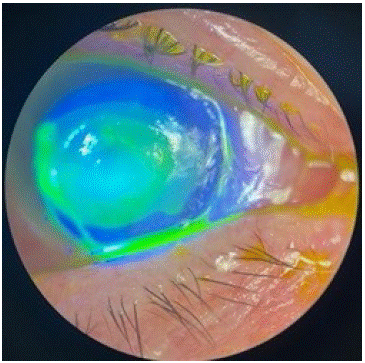
Figure 2: Patient 1 three days after initiating antibiotic treatment. Corneal ulcer fluorescein positive testing, increased in size.
The samples taken for microbiological analysis tested negative. We suspended the treatment with topical reinforced antibiotics Vancomycin and Ceftazidime due to their potential epithelial toxic effect and changed to topical Netilmicin 3 mg/ml collyrium (Netenax, Sifi group) every 6 hours to prevent infection of the ulcer. A decision was made to “heal” the ulcer, for that we added topical insulin collyrium (1IU/ml in hyaluronic acid 0.15 concentration in form of artificial tears) three times a day to accelerate the reepithelization process. Antibiotic therapy was stopped on day 7 once the appropriated regime was finished. One week after initiating treatment with topical insulin, the ulcer had been reduced by half its size (Figure 3a, 3b), and anterior chamber fibrine collection had started to clear. As the epithelization process had already begun, we added therapy with Dexamethasone phosphate collyrium (1 mg/ml) four times a day for its anti-inflammatory effect.
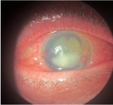
Figure 3a: Patient 1 anterior pole biomicroscopic exam 1 week after starting treatment with topical insulin (1 mg/IU) 3 times a day.
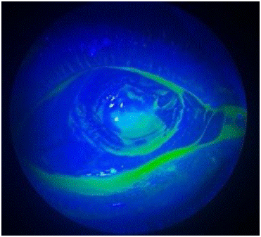
Figure 3b: Patient 1 1 week after starting treatment with topical insulin (1IU/mL) 3 times a day. Fluorescein testing positive. Ulcer reduced by half its size.
On day two after having initiated therapy with corticoids, inflammatory signs had clearly decreased, and ulcer kept diminishing in size (Figure 4a, 4b).
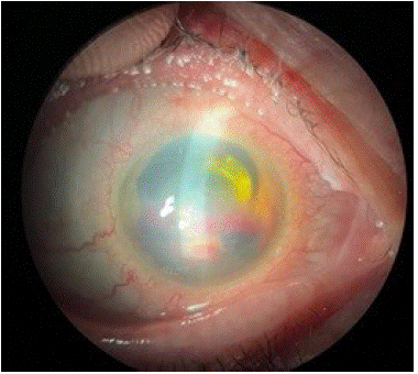
Figure 4a: Patient 1 anterior fluorescein exam on day two after starting Dexamethasone Phosphate 1 mg/mL four times a day. Reduction in size.
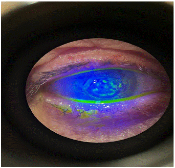
Figure 4b: Patient 1 anterior fluorescein exam on day two after starting Dexamethasone Phosphate 1 mg/mL four times a day. Reduction in size.
One week later, oral, and topical antibiotics were discontinued. Treatment with insulin collyrium 1IU/ml three times a day, Dexamethasone phosphate collyrium (1 mg/ml) four times a day, and atropine collyrium once a day were followed for one month in total. At the end of the month, cornea was mostly transparent, the ulcer had resolved, with grade I keratitis and blood adhered to the endothelium, with no fibrinous collection remaining in anterior chamber (Figure 5). After one month treatment with only topical insulin collyrium, the patient felt the air pulse at stimulation with CEB esthesiometer with 7mbar intesity.
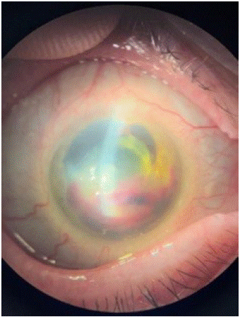
Figure 5: Patient 1 after one month treatment with Dexamethasone phosphate 1 mg/mL four times a day and Topical insuline1IU/mL three times a day., cornea mostly transparent, and blood adhered to the endothelium, with no fibrinous collection remaining in anterior chamber.
Our second patient is a male 68 years old that, also affected by type II Diabetes Mellitus, and secondary neurotrophic keratopathy type III (feeling the impulse at 7mbar intensity on Air stimulation by CEB esthesiometer). The patient came for a rutinary check-up, asymptomatic. Visual acuity for right eye was of hand movements at less than 50cm (last reported visual acuity: hand movements at 50cm). Physical exam showed intense hiperemia, a corneal ulcer fluorescein positive in inferior cornea, infiltrated and associated to fibrinous collection and hyphema in anterior chamber (Figure 6a, 6b). Corneal ulcer samples (frotis) were taken for microbiological analysis.
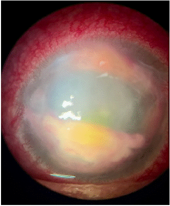
Figure 6a: Patient 2 upon admission. Anterior pole exam intense hiperemia, corneal ulcer infiltrated and associated to fibrinous collection and hyphema in anterior chamber.
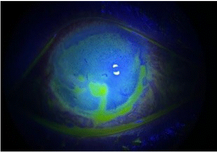
Figure 6b: Patient 2 upon admission. Fluorescein exam positive. Ulcer affecting inferior cornea.
Based on our previous experience with patient 1, we decided this not to administer intravitreous and topical reinforced antibiotics. We started this time directly with topical Netilmycin 3 mg/ml collyrium (Netenax, Sifi group) alternating every hour, oral Moxifloxacin (400 mg once a day for ten days), atropine collyrium once a day for its cycloplegic effect and added topical insulin collyrium 1IU/ml three times a day. Microbiological samples tested negative, so we decided to discontinue antibiotic use (both topical and oral) after completing 7-day treatment.
Check-ups were made every 24hours and 4 days later the ulcer had almost disappeared, with appearance of unstable aberrant epithelium (Figure 7), starting with Dexamethasone phosphate collyrium (1 mg/ml) four times a day.
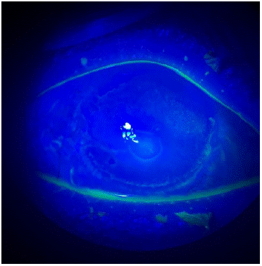
Figure 7: Patient 2 four days after treatment with topical insulin 1IU/mL 3 times a day. Fluorescein testing negative. Appearance of unstable aberrant epithelium.
Ten days after all antibiotics (topical and oral were discontinued). Four weeks later the ulcer is completely closed, no signs of infection, keratitis type I remaining, no inflammatory signs, and no fibrinous collection in anterior chamber (Figure 8a, 8b).
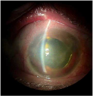
Figure 8a: Patient 2 Anterior pole exam three weeks after treatment with topical insulin 1IU/mL 3 times a day and Dexamethasone phosphate 1 mg/mL four times a day. Mild hiperemia, cornea mostly transparent, no corneal abscess, no clear hiphema or hypopyon.
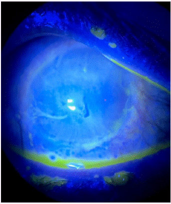
Figure 8b: Patient 2. Three weeks after treatment with topical insulin 1IU/mL 3 times a day and Dexamethasone phosphate 1 mg/mL four times a day Fluorescein test negative. Keratitis.
One month after treatment with only topical insulin collyrium and upon stimulation with CEB air esthesiometer, felt the impulse at 5mbar.
Discussion
The use of cycloplegic agents is indicated in cases of keratitis and corneal ulcers [7] with important inflammation, reducing pain and preventing from synechiae formation [7]. Our two patients presented inflammatory signs implying anterior chamber fibrinous collection in association to bleeding, that could lead to hematic Tindall. In our case we chose atropine for its longer lasting effect and because there was shortage of cyclopentolate at that time in our region. In patient one, we decided to discontinue oral acetazolamide (250 mg once a day) as it could be slowing anterior blood chamber reabsorption.
The use of anti-inflammatory therapy with topical corticosteroids at first stages infectious ulcers is controverted, existing the possibility of corneal melting, or rebound of the infection [7,8]. As a matter of that, it was of our interest to obtain a quick epithelialization of the ulcer so that we could start Dexamethasone treatment. Both patients had important signs of inflammation (including anterior chamber fibrine collection). Our patients both suffered from neurotrophic keratitis secondary to Diabetes Mellitus, which impairs normal healing of ulcers [4], and making them suitable candidates for therapy with topical insulin [6]. So, topical insulin was administered with intention of obtaining a faster epithelization process, so that corticoid therapy could be initiated as soon as possible. We saw in both cases how even before starting corticoid therapy, fibrinous reaction in anterior chamber had slowly begun to resolve. Topical Insulin’s potential anti-inflammatory defects have already been reported [5].
Both patients presented corneal ulcers, with fibrine collection in anterior chamber. Despite that, micro bacterial analysis (including bacterial, mycotic and protozoa testing) were negative. We suspect fibrine collection was inflammatory and not septic in origin, thus we did treat it from the beginning with antibiotics. As per our hospital’s protocol we treat all hypopyons with antibiotics until microbial analysis results are received. We prolonged antibiotic use for 7 more days so thar a proper regime was completed.
Although less novel, autologous plasma is another possible treatment [9] that requires patient’s blood extraction, therefore being more invasive. In our case, our patient’s had already used autologous plasma as treatment in the past and were reticent to undergo blood extractions. In addition to that, topical insulin could have a faster effect on ulcer healing than regular autologous plasma [2,6].
In literature, the principal indication of corneal topical insulin is for Persistent Corneal Epithelial Defects (PEDs) in Diabetic patients. In our day-to-day practice at our hospital, treatment of corneal ulcers with topical insulin remains an “off-label” approach, these two patients being our first cases and having signed an informed consent.
Obtaining 10ml of topical insulin collyrium requires 9 mL of hyaluronic acid 0.3% artificial tear (we chose in our case “Neovis GEL MULTI”, by Horus pharma), 1mL of physiological serum 0.9 % and 10 IU of Act rapid Insulin, all being sterile processed under a handling hood [6,9].
At this time, our patients are being treated with hyaluronic acid artificial tears and ointments exclusively. Inflammation has note reappeared, and ulcers have been completely healed, fluorescein showing keratitis punctata, probably secondary to eye dryness. We suspect neovascular formation secondary to diabetes is at the origin of anterior chamber bleeding that was associated to the fibrinous reaction, being stable right now for both patients and up to date, we have not needed to use insulin collyrium again with them.
Regarding corneal sensitivity, both patients showed clear hypoesthesia when measured with air esthesiometer (Cornea Air Esthesiometer, Brill Engines). We suspect this is due in both cases to their diabetic disease, adding the effect of scleral buckle in first patient, probably worsening the condition. However, we did not expect to see any improvement in corneal sensitivity after treatment with topical insulin, and yet we did see it. First patient, in which we suspect the effect of scleral buckle can damaged the already altered diabetic nerve bundles, didn’t feel any air pulse on stimulation at first stages, but one month after treatment with insulin collyrium, esthesiometry was of 7mbar at stimulation with Cornea Brill Esthesiometer. Second patient felt the air pulse at 7mbar first time, and one month after treatment felt it at 5mbar.
Conclusion
Corneal ulcers are a serious eye condition that require prompt diagnosis and treatment to prevent complications and vision loss, that is why it is critical to “heal” them as soon as possible, all of it avoiding excessive scarring. We think the study of corneal sensitivity is crucial for diagnosis and follow-up in diabetic patients, especially in those developing neutrophic keratopathy and secondary ulcers as was the case here. It is important in these cases to have access to a reliable esthesiometer, which can sometimes be difficult.
While traditional treatment options such as antibiotics, antifungal or antiviral medications, and corticosteroids are effective in controlling the infection and promoting healing, topical insulin may also be a promising treatment option for corneal ulcers. With further research and development, topical insulin and other novel treatment options may offer new hope for patients with corneal ulcers and improve their overall outcomes.
The use of topical insulin in treating corneal ulcers is still a relatively new and emerging treatment option, the available results of are promising and suggest that topical insulin may be a viable alternative or adjunctive treatment option for corneal ulcers.
Our ophthalmology unit is now working with the pharmacy at our hospital, selecting suitable patients, and implementing the use of this promising, non-invasive treatment.
References
- Mack HG, Fazal A, Watson S. Corneal ulcers in general practice. Aust J Gen Pract. 2022; 51: 855-860.
- Dua HS, Said DG, Messmer EM, Rolando M, Benitez-Del-Castillo JM, Hossain PN, et al. Neurotrophic keratopathy. Prog Retin Eye Res. 2018; 66: 107–31.
- Bastion ML, Ling KP. Topical insulin for healing of diabetic epithelial defects: A retrospective review of corneal debridement during vitreoretinal surgery in Malay- sian patients. Med J Malaysia. 2013; 68: 208-16.
- Bremond-Gignac D, Daruich A, Robert MP, Chiambaretta F. Recent innovations with drugs in clinical trials for neurotrophic keratitis and refractory corneal ulcers. Expert Opin Investig Drugs. 2019; 28: 1013-1020.
- Titone R, Zhu M, Robertson DM. Insulin mediates de novo nuclear accumulation of the IGF-1/insulin Hybrid Receptor in corneal epithelial cells. Sci Rep. 2018; 8: 4378.
- Diaz-Valle D, Burgos-Blasco B, Rego-Lorca D, Puebla-Garcia V, Perez-Garcia P, Benitez-Del-Castillo JM, et al. Comparison of the efficacy of topical insulin with autologous serum eye drops in persistent epithelial defects of the cornea. Acta Ophthalmol. 2022; 100: e912-e919.
- Lin A, Rhee MK, Akpek EK, Amescua G, Farid M, Garcia-Ferrer FJ, et al. Bacterial Keratitis Preferred Practice Pattern®. Ophthalmology. 2019; 126: P1-P55.
- Cabrera-Aguas M, Khoo P, Watson SL. Infectious keratitis: A review. Clin Exp Ophthalmol. 2022; 50: 543-562.
- Gregorio Maranon Hospital (no date) Hospital General Universitario Gregorio Marañón. Available at: https://www.comunidad.madrid/hospital/gregoriomaranon/sites/gregoriomaranon/files/2022-01/Insulina%201%20UIml%20colirio.pdf.