
Case Report
Austin J Clin Ophthalmol. 2024; 11(5): 1195.
Place of Mitomycin in The Treatment of Recurrent Squamous Cell Carcinoma
Z.hazil*, T.aljasser, M.Ammi, A.Jerribi , Y.Akannour, L.Seghini, E.Abdallah.
Department of Ophthalmology B, Rabat Specialty Hospital, CHU ibn Sina, Mohammed V Souissi University Rabat, Morocco
*Corresponding author: Hazil Z, Department of Ophthalmology B, Rabat Specialty Hospital, CHU ibn Sina, Mohammed V Souissi University Rabat, Morocco. Email: zahira.hazil@gmail.com
Received: September 16, 2024 Accepted: October 04, 2024 Published: October 11, 2024
Abstract
Squamous cell carcinoma is the most common malignant tumor of the bulbar conjunctiva. However, it is often underdiagnosed, leading to therapeutic delay.
We report the case of a 60-year-old patient who presented to our clinic with a rapidly progressive conjunctival tumour. Surgical excision revealed squamous cell carcinoma in situ, followed by adjuvant chemotherapy with Mitomycin-C 0.04%.
Keywords: Recurrent squamous cell carcinoma; Surgical excision; Mitomycin
Introduction
Conjunctival Squamous Cell carcinoma (CSC) is a rare malignant epithelial tumor. Conjunctival squamous cell neoplasia occurs predominantly in the elderly population; men are more often affected than women [1]; and more frequently in fair-skinned groups than in more pigmented ones [2].
Observation
This is a 60-year-old female patient with a history of prolonged sun exposure who consulted our clinic for a conjunctival growth of the left eye that rapidly increased in size over a few months and recurred after surgical excision alone on two occasions. Biomicroscopic examination of the left eye revealed a pentagonal lesion, 6 mm in long axis, located in the superior nasal bulbar conjunctiva and invading the cornea from 9 a.m. to 1 a.m. towards the visual axis, with a papillomatous appearance and several feeder vessels (Figure 1). Examination of the eyelids after eversion revealed no opposite lesions. Optical coherence tomography of the anterior segment was performed, with slices in the limbo-corneal region, but no stromal infiltration of the tumour was found. Surgical excision was performed with intraoperative application of 0.04% mitomycin (Figure 2). Pathological examination revealed a well-differentiated keratinizing squamous cell carcinoma. The patient then received 2 courses of dilute mitomycin applied locally postoperatively.
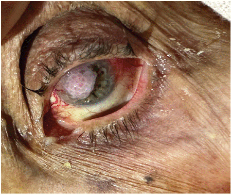
Figure 1: Photo of conjunctival lesion invading the cornea.
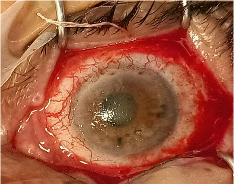
Figure 2: Post-surgical appearance after lesion exeresis.
The post-operative course was favourable, with regeneration of the tissues: normal appearance, transparent cornea, moist, transparent pink conjunctiva (Figure 3).
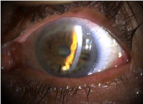
Figure 3: Slit lamp photo after 2 months of surgical treatment and mitomycin cure.
Discussion
CEC is a rare tumor, most often affecting elderly people, with a sex ratio of 1. Known risk factors for CEC are ultraviolet exposure and immunosuppression [3]. Many cases of CEC have been described in HIV-positive patients [4] or in patients undergoing long-term immunosuppressive therapy for transplantation [5].
CEC takes the form of a vegetating or budding tumor, sometimes ulcerated, with a vascular axis in each lobule [6]. It often occurs on a previously chronically irritated area (actinic keratosis and carcinoma in situ). Histology alone provides a definitive diagnosis.
The extension of CEC is primarily local, with continuity first on the surface, then deep down, making removal difficult, as in the fourth patient. Lymphatic metastases (pre-tragonal and sub-angulomaxillary) and general metastases (heart, lung, brain) are rare, and mainly concern very advanced tumours. Tumour removal must be complete. In the case of orbital invasion, exenteration is essential, along with lymph node dissection, parotidectomy and radiotherapy. The recurrence rate of ECC is high, especially in the first year following surgical excision [7].
Mytomycin C has been recommended by some in cases of recurrence, as in our patient's case, or on large tumors. However, the appearance of significant side effects (corneal ulceration, scleral necrosis, glaucoma, uveitis) means that concentrations and application times must be carefully controlled.
Khodar et al [8] apply pure Mytomycin C intraoperatively for five minutes to the scleral bed. Other authors prefer post-operative instillations of 0.02% or 0.04% Mitomycin C [9].
Conclusion
Les traitements actuels ne permettent pas d’éliminer les récidives fréquentes du CEC. Aussi un suivi régulier des patients apparaît essentiel. Le traitement est chirurgicale soigneuse. L’étude anatomopathologique de la pièce est la seule à garantir le diagnostic de certitude. Il reste à sensibiliser la population aux facteurs de risque et à l’importance de la protection contre les rayons ultraviolets.
Author Statements
Conflicts of Interest
All authors declare that they have no conflicts of interest.
References
- Amoli FA, Heidari AB. Survey of 447 patients with conjunctival neoplastic lesions in Farabi Eye Hospital, Tehran, Iran. Ophthalmic Epidemiol. 2006; 13: 275-9.
- Shields CL, Demirci H, Karatza E, Shields JA. Clinical survey of 1643 melanocyticand nonmelanocytic conjunctival tumors. Ophthalmology. 2004; 111: 1747-54.
- D Acis, A Donnio, L Ayéboua, R Richer, J Guyomarch, A Warter, et al. Carcinome épidermoïde conjonctival.à propos de quatre cas aux Antilles JFO-05/2008. Serviced’Ophtalmologie, Centre Hospitalier Universitaire de Fort deFrance, Hôpital Pierre Zobda-Quitman.
- Weinstein JE, Karp CL. Ocular surface neoplasias andhuman immunodeficiency virus infection. Curr Opin Infect Dis. 2013; 26: 58-65.
- Abdallah ES, Caml LS, Shields JA, Ralph MD. Eagle, Phila-delphia, Pa aggressive coniunctival squamous cell carcinomain a patient following liver transplantation. Arch Ophthalmol. 2003; 121: 280-2.
- Elmaleh C, D’Hermies F, Schwartz L, Renard G, Pouliquen Y. Carcinome invasif du limbe conjonctivo-cornéen. J Fr Ophtalmol. 1993; 16: 417-9.
- Tabin G, Levin S, Snibson G, Loughnan M, Taylor H. Late recurrences and the necessity for long-term follow-up in corneal and conjunctival intraepithelial neoplasia. Ophthalmology. 1997; 104: 485-92.
- hokhard S, Soni A, Singh Sethi H, Sudan R, Sony P, Pangtey MS. Combined surgery, cryotherapy, and mitomycin-C for re current ocular surface squamous neoplasia. Cornea. 2002; 21: 189-91.
- fferson T. Topical mitomycin C for extensive, recurrent conjunctivalcornea lsquamous cell carcinoma. Am J Ophthalmol. 2003; 135: 122-3.