
Case Report
Austin J Clin Ophthalmol. 2024; 11(5): 1197.
Unique Presentation of Focal Choroidal Excavation with Solitary Congenital Hypertrophy of the Retinal Pigment Epithelium
Omar N Mehana, MSc*; Haaris A Shiwani, FRCOphth; Usman K Hayat, FRCOphth; Guillermo De La Mata, FEBO, Ali A Lamin, PhD
Manchester Royal Eye Hospital, Manchester Foundation Trust, United Kingdom
*Corresponding author: Omar Mehana, Department of Ophthalmology, Manchester Royal Eye Hospital, Manchester, M13 9WL, United Kingdom. Tel: +447541369385 Email: Omar.Mehana@mft.nhs.uk
Received: November 07, 2024; Accepted: December 02, 2024; Published: December 09, 2024
Abstract
Congenital Hypertrophy of the Retinal Pigment Epithelium (CHRPE) is a rare benign, flat, hyperpigmented lesions arising from Retinal Pigmented Epithelium (RPE) & Focal Choroidal Excavation (FCE) is a concave choroidal excavation typically located in macular area & normally identified by Optical Coherence Tomography (OCT).
This case report aims to present & explore characteristic features of both CHRPE & FCE, to identify different variant, possible associations & to emphasise the importance of imaging in identifying and monitoring these lesions.
Keywords: Focal Choroidal Excavation; CHRPE
Introduction
CHRPE and FCE represent two unique and distinct entities that can affect the retina. CHRPE typically manifests as a rare, benign and asymptomatic hyperpigmented lesions on the fundus [1]. These lesions are categorized into three main forms: solid (unifocal), grouped (multifocal), and atypical [1]. A typical (unifocal) CHRPE presents as a single, flat, round lesion with smooth, sharply defined borders and can range in colour from light brown or grey to black [2]. These typical lesions are characteristically surrounded by a narrow hypopigmented halo [2]. CHRPE often has lacunae present which is an atrophied window-like defect, these lacunae can vary in size shape and abundance [3]. They are not however predictors of serious pathology but rather an intersubject variable [3]. Typical (multifocal) are a group of lesions arranged in clusters usually in one quadrant of the retina [4]. These lesions are termed “bear tracks” in the literature due to their animal footprints resemblance [3,4]. Atypical CHRPEs are often bilateral and may spread across the entire fundus. This variant is sometimes associated with Familial Adenomatous Polyposis Coli (FAPC) or Gardner's syndrome [4].
FCE is identified as a concave area in the choroid, typically located in the macular region, though it can occasionally appear outside this area [5]. FCE is usually detected via OCT and does not involve accompanying scleral ectasia or posterior staphyloma [5]. It predominantly occurs in individuals in their fourth or fifth decade of life, is generally unilateral, and shows no sex predilection [5]. FCEs are not inherently progressive and may occur in any quadrant of the fundus, though slight predilection observed in the temporal quadrant [5].
On examination, FCE may present as mild pigmentary changes or hypopigmented lesions, primarily in the macular area, though they are not always visible clinically. OCT imaging reveals a concavity in the choroid with the RPE following the contour of this concavity. FCEs are classified into two types: conforming and non-conforming. Conforming FCEs have no separation between the photoreceptor layer and the underlying RPE, whereas non-conforming FCEs display a hyporeflective space between these layers, sometimes containing hyperreflective material, which may indicate inflammatory substances or degenerated outer segment residue depending on the cause [5]. FCE can be further classified into either congenital (primary) or acquired (secondary) [5].
We present a rare case of the presence of FCE within a CHRPE in a middle-aged female patient.
Case Presentation
A 43-year-old female was referred to the medical retina assessment clinic for a review and fundus examination following an incidental finding in the left eye, identified by a local optometrist. The patient reported mild symptoms, including dry eyes and occasional headaches, likely due to work-related, prolonged, computer use. Her personal and family medical history was unremarkable, and she reported no family history of any polyps or colonic cancer. The bestcorrected visual acuity in both eyes was -0.1 LogMAR (6/4.8 Snellen visual acuity), the anterior segment examination was unremarkable, and intraocular pressure was within normal limits.
Fundus examination of the left eye revealed a well-demarcated, round, pigmented lesion located below the inferior retinal vessels at the inferior arcade, measuring 2.2 mm by 2.5 mm. Additionally, there was a faint amelanotic halo on the superior margin and a subtle pigmented halo surrounding this amelanotic halo. FAF showed absolute hypo-autofluorescence. OCT revealed an excavated lesion with irregular RPE, thinning of the outer retinal layers, intraretinal cystic changes, and a conforming FCE. Choroidal thinning was also noted at the base of the excavation. The examination of the right eye was unremarkable. No intervention was indicated, and the patient was put on a routine monitoring pathway.
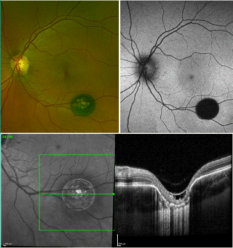
    Figure 1: Imaging of a 43-year-old female patient with solitary CHRPE and
FCE in the left eye. A) Colour fundus photograph of the left eye demonstrating
a well-demarcated, round, pigmented lesion located below the inferior retinal
vessels at the inferior arcade, measuring 2.2 mm by 2.5 mm with a faint
amelanotic halo on the superior margin and a subtle pigmented halo on the
outer margin of this. B) Fundus autofluorescence demonstrating a round
well demarcated absolute hypo-auto fluorescent lesion correlating to colour
fundus photograph. C) OCT of left eye showing an excavated lesion with
irregular RPE, thinning of the outer retinal layers, intraretinal cystic changes,
and a conforming FCE with choroidal thinning at the base of the excavation.
Discussion
CHRPE and FCE are generally considered distinct conditions and are not known to co-occur. Solitary CHRPE is congenital and typically benign, while atypical CHRPE as mentioned previously can be associated with FAPC. FCE may be either congenital or acquired, with some acquired cases linked to inflammation, pachychoroid spectrum disease and retinal dystrophy. FCE has also been associated with Multiple Evanescent White Dot Syndrome (MEWDS), a rare form of posterior uveitis, as well as myopia, although FCE is relatively uncommon in myopic individuals [5].
Shields et al. [6] analysed the clinical characteristics of solitary CHRPE in a study involving 335 eyes of 330 patients. Their findings indicated that these lesions were most commonly found in the inferotemporal quadrant (31%) and the equatorial region (45%). They were rarely located in the macula (1%) or the peripapillary region (1%). In 325 patients (98%), the lesions were unilateral, and in 297 patients (88%), the lesions were darkly pigmented. All the lesions were well demarcated (100%) and nearly all were completely flat (99%). The size of these lesions varied from 0.2 mm to 13 mm. The margins of the lesions were regular in 211 patients (63%) and irregular in 126 patients (37%). Additionally, a pigmented halo was present in 193 patients (57%), while a nonpigmented halo was noted in 154 patients (46%). The description in this pivotal study supports the classification of the pigmented lesion in our case as a solitary CHRPE.
In a study by Shinojima et al. [7], FCE was associated with various choroidal diseases, including Polypoidal Choroidal Vasculopathy (PCV), idiopathic FCE, central serous Chorioretinopathy (CSCR), wet Age-Related Macular Degeneration (ARMD), and idiopathic Choroidal Neovascular Membrane (CNVM). Lee et al. [8] found that CSCR and CNVM were frequently associated with FCE, with 24% of eyes having CSCR and 22% CNVM. CSCR is one of the most documented associations with FCE.
In a study by Ellabban et al. [9], FCE was identified in 7.8% of 116 consecutive eyes with CSCR. These patients often present with active CSC and resulting vision loss. Type 1 and 2 CNVMs have been reported to occur around choroidal excavation, with type 2 CNVMs potentially obscuring the underlying FCE, complicating accurate diagnosis. Patients with CNVM typically have poorer visual outcomes compared to those with CSCR. Xu et al. [10] reported that out of 15 cases of FCE, 12 had CNVM at presentation, and 3 developed CNVM during follow-up. The literature describes only a single case of a combined presentation of FCE and CHRPE.
Üçer et al [1] have presented a similar case to that which we have described above, a 46-year-old female who presented with a focal choroidal excavation in association with solitary CHRPE. Both patients were females in the same age group. The lesions were both well-demarcated, round, flat, darkly pigmented areas and relatively near in size. The OCT findings were similar, showing irregular RPE, thinning of the outer retinal layer and choroidal thinning at the base of excavation. The case presented by Üçer et al [1] was however a nonconforming FCE with a more superficial excavation.
While solitary CHRPE and FCE are generally benign conditions, it is crucial to differentiate this unique entity from conditions such as choroidal melanoma, choroidal nevus, melanocytoma, focal pigmentation, or congenital toxoplasmosis. This differentiation is important to ensure appropriate follow-up and management.
This case emphasizes the fundamental role of multimodal imaging including colour fundus photography and FAF in documenting and monitoring the appearance, size and characteristics of the lesion. OCT is a fundamental imaging technique to look at the lesions and aid in diagnosis. It provides better visualisation of retinal layers and choroid. OCT is essential in diagnosing FCE. Fluorescein Angiography and Indocyanine Green Angiography have a role in identifying or ruling out CNVM and visualising the choroidal vasculature. Once CHRPE and FCE are confirmed and secondary causes are excluded, it is deemed safe to monitor these patients in primary or secondary care settings.
Conclusion
CHRPE and FCE are benign, distinct rare conditions. However, it is important to be cognisant of the potential for co-presentation. Accurate identification of these conditions is fundamental for appropriate monitoring and follow-up as they can mimic more sinister ophthalmic pathology. Multimodal imaging plays a fundamental role in identifying pathology and in monitoring changes.
Author Statements
Conflict of Interest
The authors declare no conflicts of interest.
Funding
This study received no funding.
Author Contributions
Omar Mehana and Ali Lamin had full access to the data and take responsibility for the integrity of the data and that the manuscript is not under consideration for publication elsewhere.
References
- Üçer MB. Focal choroidal excavation in solitary congenital hypertrophy of the retinal pigment epithelium. J Fr Ophtalmol. 2024; 47: 104123.
- Buettner H. Congenital hypertrophy of the retinal pigment epithelium. Am J Ophthalmol. 1975; 79: 177-89.
- Coleman P, Barnard NAS. Congenital hypertrophy of the retinal pigment epithelium: prevalence and ocular features in the optometric population. Ophthalmic Physiol Opt. 2007; 27: 547-55.
- Ireland AC, Rodman J. Congenital Hypertrophy of Retinal Pigment Epithelium. 2024 May 15. In: StatPearls. Treasure Island (FL): StatPearls Publishing. 2024.
- Verma S, Kumar V, Azad S, Bhayana AA, Surve A, Kumar S, et al. Focal choroidal excavation: review of literature. Br J Ophthalmol. 2021; 105: 1043- 1048.
- Shields CL, Mashayekhi A, Ho T, Cater J, Shields JA. Solitary congenital hypertrophy of the retinal pigment epithelium: clinical features and frequency of enlargement in 330 patients. Ophthalmology. 2003; 110: 1968-76.
- Shinojima A, Kawamura A, Mori R, Yuzawa M. Morphologic features of focal choroidal excavation on spectral domain optical coherence tomography with simultaneous angiography. Retina. 2014; 34: 1407-14.
- Lee CS, Woo SJ, Kim YK, Hwang DJ, Kang HM, Kim H, et al. Clinical and spectral-domain optical coherence tomography findings in patients with focal choroidal excavation. Ophthalmology. 2014; 121: 1029-35.
- Ellabban AA, Tsujikawa A, Ooto S, Yamashiro K, Oishi A, Nakata I, et al. Focal choroidal excavation in eyes with central serous chorioretinopathy. Am J Ophthalmol. 2013; 156: 673-83.
- Xu H, Zeng F, Shi D, Sun X, Chen X, Bai Y. Focal choroidal excavation complicated by choroidal neovascularization. Ophthalmology. 2014; 121: 246-250.
- Meyer CH, Holz FG. Documentation of congenital hypertrophy of the retinal pigment epithelium with wide-field funduscopy. Semin Ophthalmol. 2009; 24: 251-3.
- Margolis R, Mukkamala SK, Jampol LM, Spaide RF, Ober MD, Sorenson JA, et al. The expanded spectrum of focal choroidal excavation. Arch Ophthalmol. 2011; 129: 1320-5.