Clinical Image
Austin J Clin Ophthalmol. 2025; 12(1): 1199.
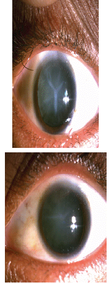
Sutural Cataract - Clinical Picture
Zeinebou H’meimett*
Clinique Ridwane, Nouakchott, Mauritanie
*Corresponding author: Zeinebou H’meimett, Clinique Ridwane, Nouakchott, Mauritanie Email: zeinebou_h@yahoo.com
Received: June 11, 2025 Accepted: June 16, 2025 Published: July 18, 2025
Clinical Image
We report the case of a 45-year-old female patient with no particular medical history who presented with a progressive decline in visual acuity in both eyes since birth. Her best correction was 2/10 in the right eye and 8/10 in the left. Ocular tone was normal. Biomicroscopic examination revealed a Y-shaped sutural cataract on the right eye with a slight progressive posterior subcapsular lesion on the left. Fundus examination was normal.

The patient underwent cataract surgery by phacoemulsification in the right eye, which was uneventful, with an uneventful postoperative course and recovery to 9/10 with correction. Regular follow-up was performed to monitor the progression of the cataract in the left eye and to detect a complication in the right eye. The impact of congenital cataracts on visual acuity varies depending on the type of cataract. This congenital cataract follows the embryonic suture lines of the lens.