
Clinical Image
Austin J Clin Ophthalmol. 2025; 12(1): 1200.
Prepapillary Arterial Loop Associated with A Branch Retinal Artery Occlusion
Achegri Y*, Bouimtarhan A, Azib S, Bouabadi C, Fiqhi A, El Khoyaali A and Mouzari Y
Department of Ophtalmology, Mohamed V Military Study Hospital Rabat, Morocco
*Corresponding author: Achegri Y, Department of Ophtalmology, Mohamed V Military Study Hospital Rabat, Morocco Email : r.achegriyoussef@gmail.com
Received: August 09, 2025 Accepted: September 08, 2025 Published: September 10, 2025
Clinical Image
Fundus photography of the left eye of a 45-year-old female patient demonstrating features consistent with a branch retinal artery occlusion (BRAO) involving the superior temporal quadrant (Figure 1). The affected retinal territory exhibits well-demarcated retinal whitening and edema, corresponding to ischemia of the inner retinal layers. The foveal region appears partially involved, causing symptomatically a decrease in visual acuity. A distinct prepapillary vascular loop is observed, originating from the optic nerve head, projecting anteriorly into the vitreous cavity, and re-entering the retinal surface before continuing as a branch artery. No associated neovascularization, hemorrhage, or optic disc edema is evident. The fundus photography of the right eye shows a same distinct prepapillary vascular loop, without arterial occulsion (Figure 2).
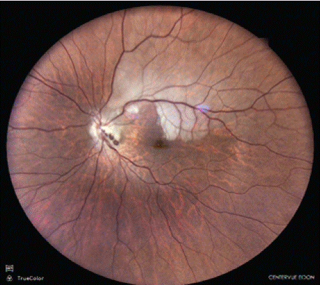
Figure 1:
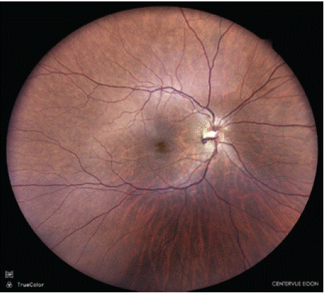
Figure 2: