
Case Report
Austin J Clin Ophthalmol. 2025; 12(1): 1201.
Fundus Flavimaculatus Complicated by Massive Subchoroidal Hemorrhage
Elkhoyaali A*, Laaouina S, Ouraoumo F, Zerrouk R, Fiqhi A and Mouzarii Y
Ophthalmology Department, Mohammed V Military Teaching Hospital, Rabat, Mohammed V University, Hay Ryad, 10100 Rabat, Morocco
*Corresponding author: Elkhoyaali Adil, Ophthalmology Department, Mohammed V Military Teaching Hospital, Rabat, Mohammed V University, Hay Ryad, 10100 Rabat, Morocco Email: Elkhoyaaliadil@gmail.com
Received: August 19, 2025 Accepted: September 09, 2025 Published: September 11, 2025
Abstract
Fundus flavimaculatus (FFM) is an entity of Stargart’s disease defined by Franceschetti as a hereditary, bilateral retinal dystrophy characterized by the presence of poorly defined, pisciform or radial yellowish spots located in the pigment epithelium (PE) and called flecks. It is the most common form of juvenile macular dystrophy, representing 7% of all retinal dystrophies. More or less severe forms can be distinguished depending on the age of onset of functional signs and their association or not with areas of macular atrophy. The progression is towards atrophy in the short or long term. Neovascular complications have rarely been described in this dystrophy. Some authors have reported the efficacy of intravitreal injections of anti-VEGF (vascular endothelial growth factor) in the treatment of these neovascularizations. We report an extremely rare case of a 56-year-old Moroccan woman who presented with a sudden visual acuity loss related to a massive subchoroidal hemorrhage complicating choroidal neovascularization in the right eye on a bilateral fundus flavimaculatus on fundus examination. Our patient presented with classic features of FFM complicated by choroidal neovascularization; clinically, on autofluorescence imaging, and on angiography. The evolution was favorable eight weeks after an intravitreal injection of anti-VEGF; vision improved with a reduction in subretinal fluid as well as partial resorption of the hemorrhage and retraction of the neovascularization on fundus and optical coherence tomography.
Keywords: Fundus flavimaculatus; Choroidal neovascularization; ANTI VEGF
Introduction
Fundus flavimaculatus (FFM) is a rare form of Stargardt syndrome, with whitish yellow lesions that resemble either the tail of a fish or round spots on the retina. It primarily affects young patients, who ultimately suffer visual loss associated with atrophy or flecks in the macular area within a short or long-time frame. This condition may also be complicated by choroidal neovascularization (CNV) [1- 4].
We report a very rare case of FFM with CNV and a significant subchoroidal hemorrhage, that also exhibited a remarkable response to anti-VEGF treatment.
Observation
We present a case of a 56-year-old female patient with late identified FFM, who is on hemodialysis and receiving heparin for end-stage renal failure with a past medical history of idiopathic membranoproliferative glomerulonephritis. The patient presented with acute left-eye (LE) vision loss without pain or redness.
On clinical examination, her vision was severely reduced in the LE to hand motion only; she had preserved vision in the right eye (RE) to 10/10. Both eyes had relatively normal anterior segments and intraocular pressure. Fundus exam revealed multiple yellowishwhite flecks, predominantly pisciform but with a few round ones, distributed in the macular region (Figure 1). There were signs of arteriovenous crossing and there was also dense retrohyaloid and subretinal hemorrhage, covering the entire macular area in the LE (Figure 2).
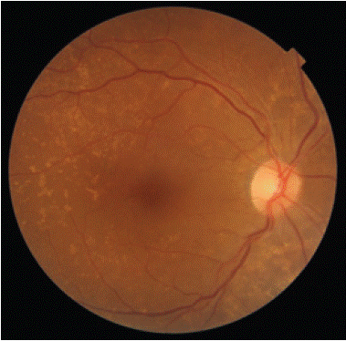
    Figure 1: Fundus right eye: multiple yellowish-white flecks, predominantly
pisciform but with a few round ones, distributed in the macular region.
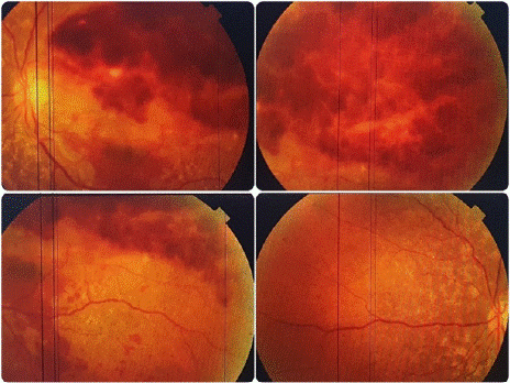
    Figure 2: Fundus exam left eye: dense retrohyaloid and subretinal
hemorrhage, covering the entire macular area in the LE.
Autofluoroscence imaging revealed multiple hyperautofluorescent lesions (44 of the hemorrhagic zone) corresponding to the flecks. Fluorescein angiography of the LE showed hypofluorescence caused by decreased transmission due to the presence of hemorrhage and superior hyperfluorescence with leakage possibly indicating neovascularization. In the RE, hyperfluorescence focally at the fleck sites corresponded to pigment epithelial changes (Figure 3).
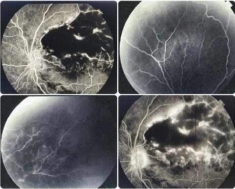
Figure 3: Fluorescein angiography of the LE.
In the left eye, the findings on OCT angiography were obscured by hemorrhage and did not allow clear CNV demonstration. The spectral domain OCT (SD-OCT) of the right eye did show juxtafoveal CNV with associated subretinal fluid and hyperreflective dots (Figure 4). B-wave echo of the left eye shows extensive subretinal hemorrhage (Figure 5)
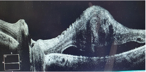
    Figure 4: The spectral domain OCT (SD-OCT) of the right eye did show
juxtafoveal CNV with associated subretinal fluid and hyperreflective dots.
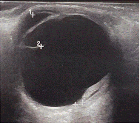
    Figure 5: B-wave echo of the left eye shows extensive subretinal
hemorrhage.
Thus, based on the findings, we made the diagnosis of FFM with CNV, left eye.
Therefore, we adjusted the heparin therapy. The patient received a single intravitreal injection of anti-VEGF in his left eye. At 10-weeks of follow-up, there was partial hemorrhagic resorption and improvement of visual acuity to 4/10 (Figure 6), and no metamorphopsia. The OCT showed minimal subretinal fluid, regressing CNV, and adjacent scarring (the subretinal exudation resolved with residual scar on the right eye).
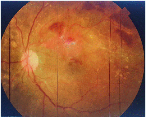
Figure 6: Fundus exam: partial hemorrhagic resorption.
Discussion
Franceschetti introduced the term “fundus flavimaculatus” in 1963 to refer to yellowish flecks that are fusiform, round or fish shaped, distributed throughout the posterior pole and into the midperiphery with the onset of central vision loss [5].
Today, FFM and Stargardt disease are considered to belong to the same spectrum [6]. FFM is defined by lesions occurring in the peripheral vision and later in life, whereas Stargardt dystrophy is characterized by lesions that are more centralized and part of an early onset [7]. Both conditions are classically diagnosed with autosomal recessive inheritance (~90%), although autosomal dominant forms exist [8-9].
There are few histopathological reports. Klein and Krill provided histological evidence of the deposition of mucopolysaccharides, and Eagle et al. have reported evidence of accumulation of lipofuscin in the retinal pigment epithelium and subsequent ruptures of Bruch's membrane leading to CNV [3,10-12].
Foveal preservation can sustain normal vision for some time [6], but progressive macular atrophy is seen in 90% of occurrences and CNV is rare [2–4, 6]. Visual symptoms usually begin at 30-40 years of age and are slow to progress [13,14].
Autofluorescence is a signature symptom of FFM. Lois et al. described variable autofluorescence with the peripapillary sparing in younger patients [15]. Abnormal electroretinograms (ERGs) are consistent features [16]. The fluorescein angiography demonstrates, flecked lesions with early hypofluorescence from masking and the hyperfluorescent borders being changes in adjacent epithelium. Although it is rare to see CNV in FFM, it usually arises close to atrophic zones in patients over the age of 50 [2–4, 17]. It may be mistaken for age related macular degeneration (AMD) if the flecks are misinterpreted as drusen [18].
The therapeutic modalities for choroidal neovascularization (CNV) in FFM remain poorly defined, and treatment options include observation [2–4,19], laser photocoagulation [4, 20], and photodynamic therapy (PDT) [21], and intravitreal triamcinolone and anti-VEGF agents. There are many treatment options, but the choice depends on the size and location of the CNV relative to the macula. Although Souied et al. reported positive results following the use of photodynamic therapy in small case series [6,18,22], there was still a chance of long-term damage to the retinal pigment epithelium. Macular translocation has also been reported by some authors as a treatment option [23].
A number of publications have reported excellent visual outcomes following intravitreal anti-VEGF injections, most recently from 1 to 3 injections overall [24-26].
In our case, the recommendation for anti-VEGF therapy was based on considerable vision loss from massive hemorrhage. The patient's concurrent anticoagulant therapy while on dialysis further complicates management, where the preservation of the visual prognosis is not the primary concern. Therefore, regular angiographic monitoring of the fellow eye is needed. Remarkably, the CNV convalesced completely after a single intravitreal injection with no recurrence at one year post-injection.
Conclusion
Fundus flavimaculatus is an uncommon condition with a poor understanding and can lead to visual loss in young individuals, and in some rare cases, further complications like choroidal neovascularization, and hemorrhage. This case demonstrates the utility of intravitreal anti-VEGF injections.
References
- Coscas G, Gaudric A, Barthelemy F. Un cas de fundus flavimaculatus avec neovaisseaux preretiniens. J Fr Ophtalmol. 1980; 3: 27- 32.
- Bottoni F, Fatigati G, Carlevaro G, De Molfetta V. Fundus flavimaculatus and subretinal neovascularization. Graefes Arch Clin Exp Ophthalmol. 1992; 230: 498-500.
- Leveille AS, Morse PH, Burch JV. Fundus flavimaculatus and subretinal neovascularization. Ann Ophthalmol. 1982; 14: 331-334.
- Klein R, Lewis RA, Meyers SM, Myers FL. Subretinal neovascularization associated with fundus flavimaculatus. Arch Ophthalmol. 1978; 96: 2054- 2057.
- Franceschetti A, Francois J. Fundus flavimaculatus. Arch Ophtal- mol. 1965; 25: 505-530.
- Valmaggia C, Niederberger H, Helbig H. Photodynamic therapy for choroidal neaovascularization in fundus flavimaculatus. Retina. 2002; 22: 111-113.
- Roy R, Kumar S, Chandrasekharan DP, Ghose A, Sharma P. Multimodal imaging in multifocal pattern dystrophy simulating fundus flavimaculatus. Indian J Ophthalmol. 2016; 64: 395-396.
- Bird AC. Retinal photoreceptor dystrophies. Am J Ophthal- mol. 1995; 119: 543-562.
- Donoso LA, Edwards AO, Frost A, Vrabec T, Stone EM, Hageman GS, et al. Autosomal dominant Stargardt-like macular dystrophy. Surv Ophthalmol. 2001; 46: 149-163.
- Klein BA, Krill AE. Fundus flavimaculatus. Clinical, func- tional and histopathological observations. Am J Ophthal- mol. 1967; 64: 3-23.
- Eagle RC, Lucier AC, Bernaddino VB, Yanoff M. Retinal pigment epithelial abnormalities in fundus flavimaculatus. Ophthalmology. 1980; 87: 1189-1200.
- Battaglia Parodi M, Da Pozzo S, Ravalico G. Photodynamic therapy for choroidal neovascularization associated with pattern dystrophy. Retina. 2003; 23: 171-176.
- T. M. Aaberg, “Stargardt’s disease and fundus flavimaculatus: evaluation of morphologic progression and intrafamilial co- existence,” Transactions of the American Ophthalmological Soci- ety. 1986; 84: 453–487.
- J. D. Armstrong, D. Meyer, S. Xu, and J. L. Elfervig, “Long-term follow-up of Stargardt’s disease and fundus flavimaculatus,” Ophthalmology. 1998; 105: 448–457.
- Lois N, Halfyard AS, Bird AC, et al. Fundus autofluorescence in Stargardt macular dystrophy-fundus flavimaculatus. Am J Ophthalmol. 2004; 138: 55- 63.
- Lois N, Holder GE, Bunce C, et al. Phenotypic subtypes of Stargardt macular dystrophy-fundus flavimaculatus. Arch Ophthalmol. 2001; 119: 359-369.
- Gass JDM, Hummer J. Focal retinal pigment epithelial dysplasia associated with fundus flavimaculatus. Retina. 1999; 19: 297-301.
- Souied EH, Pawlak D, Algan M, et al. Photodynamic therapy for choroidal neovascularization on late-onset fundus flavimaculatus. Am J Ophthalmol. 2005; 140: 312-314.
- R. Klein, R. A. Lewis, S. M. Meyers, and F. L. Myers, “Subreti- nal neovascularization associated with fundus flavimaculatus,” Archives of Ophthalmology. 1978; 96: 2054–2057.
- D. Pawlak, E. Souied, G. Mimoun, M. Papp-Pawlak, G. Coscas, and G. Soubrane, “Fundus flavimaculatus and choroidal neo- vascularization,” Journal Francais d’Ophtalmologie. 2006; 29: 188–194.
- B. Braun, U. Schneider, P. Hasler, and C. Pru nte, “Combination therapy of PDT and triamcinolone in CNV associated with fundus flavimaculatus,” Klinische Monatsblatter fur Augen- heilkunde. 2007; 224: 353–355.
- Pawlak D, Glacet-Bernard A, Papp M, Coscas G, Soubrane G. Resultats de la translocation maculaire limitee dans la neovascula- risation choroidienne retrofoveale de la degenerescence maculaire liee a l’age. J Fr Opthalmol. 2004; 27: 3S31-37.
- Gass JDM. Stereoscopic Atlas of Macular Diseases. Ed 2. St Louis, CV Mosby Co, 1977: 40-77.
- N. Tejerina, M. Garc ia-Pous, E. P. Pozos, M. C. D. Esteban, and J. M. Boronat. “Ranibizumab for choroidal neovasculariza- tion in fundus flavimaculatus,” Retinal Cases & Brief Reports. 2008; 2: 250–252.
- C. Quijano, G. Querques, N. Massamba, G. Soubrane, and E.H. Souied. “Type 3 choroidal neovascularization associated with fundus flavimaculatus,” Ophthalmic Research. 2009; 42: 152–154.
- V. Koh, T. Naing, and C. Chee. “Fundus flavimaculatus and choroidal neovascularization in a young patient with normal electroretinography: case report,” Canadian Journal of Ophthal- mology. 2012; 47: e3–e5.