Case Report
Austin J Clin Ophthalmol. 2015;2(2): 1043.
Radiation Therapy for Eyelid Basal Cell Carcinoma - A Case Report
Cardoso MS1*, Monteiro SG1, Pires J1, Mariano M1, Castela G2, Marques RV3 and Lopes N1
1Department of Ophthalmology, Centro Hospitalar do Baixo Vouga, Portugal
2Department of Ophthalmology, Centro Hospitalar e Universitàrio de Coimbra, Portugal
3Department of Radiation Therapy, Portuguese Institute of Oncology Francisco Gentil Coimbra, Portugal
*Corresponding author: Cardoso MS, Department of Ophthalmology, Centro Hospitalar do Baixo Vouga - Hospital Infante D. Pedro, Avenida Artur Ravara, 3814-501 Aveiro
Received: January 20, 2015; Accepted: February 26, 2015; Published: February 27, 2015
Abstract
Basal cell carcinoma is the most common type of eyelid cancer. Although rarely associated with metastatic disease, it is locally invasive and can cause significant destruction and cosmetic deformities. The first line treatment is usually surgical excision, but currently numerous alternatives are available; the radiation therapy is one of these therapeutic options.
A case report of an eyelid basal cell carcinoma with intraorbital extension successfully treated with radiation therapy, in a female patient with 87 years of age, is presented. Complementary tests results are described, as well as clinical evolution.
We concluded that radiation therapy is a good treatment option for unresectable tumors and for patients who are unable to tolerate more invasive methods.
Keywords: Basal cell carcinoma; Eyelid; Orbit; Treatment; Radiation therapy
Introduction
Basal Cell Carcinoma (BCC) is a slow-growing and locally invasive skin tumor which rarely metastasizes, derived from cells of the basal layer of the epidermis [1]. It is the most common type of cancer in Europe, Australia and the USA, affecting predominantly individuals of Caucasian race and older than 60 years [2-4]. The most significant risk factors appear to be exposure to ultraviolet radiation and genetic predisposition [5]. It usually occurs on sun-exposed areas of skin, and therefore about 74% of cases arise in the head and neck [6]. Although relatively indolent, this tumor can be quite destructive to cause significant cosmetic deformities [7]. Currently, there are several treatment modalities for BCC, such as surgical excision, radiotherapy, topical 5% imiquimod cream, cryotherapy and photodynamic therapy [8]. The therapeutic choice will depend on many factors, including the histologic type of tumor, its size and location, patient's age, general health and expectations, treatment availability and also in the experience and preferences of the specialist [9]. The first line treatment is usually surgical excision, showing the lowest failure rates [10]. However, radiation therapy is a very effective treatment strategy, particularly for patients with primary lesions requiring difficult or extensive surgery [11,12]. The aims of any therapy selected for BCC treatment are to ensure complete removal, the preservation of function and a good cosmetic outcome [4].
Case Report
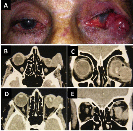
An 87-year-old Caucasian woman presented with an extensive mass (25 mm in its major axis) located in the temporal portion of the left lower eyelid that had been slowly growing for the last two years (Figure 1A). As ophthalmologic antecedents, she had phthisis bulbi in her left eye caused by glaucoma. The histopathological analysis of the lesion revealed a basal cell carcinoma without lymphatic or vascular invasion. Computed Tomography (CT) of the orbits showed a well-circumscribed mass in the infratemporal region of the left orbit with intraorbital extension (Figures 1B, 1C, 1D and 1E). The tumor was classified as T3N0M0.

Figure 1: A. Macroscopic aspect of the CBC the left lower eyelid. B-E. Orbital CT scan showed a well-circumscribed mass in the infratemporal region of the left orbit with intraorbital extension.
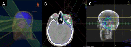
Due to the advanced age of the patient, the considerable size of the tumor, its difficult-to-access anatomical location and the irreversible status of her left eye we opted for treatment with radiation therapy. The patient was then referred to the Portuguese Institute of Oncology of Coimbra, where she was subjected to three-dimensional conformal external beam radiation therapy (Figure 2A, 2B and 2C). The patient received a total dose of 30 Gray (Gy) divided into 10 fractions over a 2-week period. The treatment was well tolerated and there were no serious adverse events.

Figure 2: A. Three-dimensional representation of the radiation beams used in the treatment. B. Radiation therapy fields, isodose curves (%), volumes to radiate and organs at risk. C. Projection of the fields of treatment, with multileaf protection.
Two months after treatment, a significant improvement was found with complete tumor regression confirmed by CT-scan (Figure 3A).
After 2 years of follow up, there has been no complication or recurrence of the lesion and the aesthetic outcome was excellent (Figure 3B).

Figure 3: A. Orbital CT scan after treatment with external beam radiation therapy, showing complete tumor regression. B. Macroscopic appearance after treatment with external beam radiotherapy.
Discussion
The goal of treatment of eyelid BBC is the complete removal of tumor cells, with preservation of the unaffected eyelid and periorbital tissues. Although there are several nonsurgical therapeutic approaches available, surgery is generally considered the treatment of choice for this type of tumor [13]. It is recommended a surgical resection margin of about 3-4 mm. The Mohs micrographic surgery has the highest cure rate of any procedure and it also allows the greatest preservation of normal tissue [14]. In this technique, the excised tissue is frozen and sectioned horizontally, providing a three-dimensional mapping of the tumor; the layers are then examined histologically during the intraoperative period, in order to obtain free margins of tumor [4].
Radiation therapy plays an important role in the treatment of benign and malignant orbital disease [15]. It is a good therapeutic option for tumors whose location and size determine total surgical excision and in patients who are unwilling or unable to tolerate more invasive methods [9,13].
The use of external beam radiation therapy for eyelid malignancies results in local control in 93.3-96.5% of cases [13,16]. Despite these promising results, a long-term follow-up is needed due to the risk of recurrence [16]. A randomized trial by Avril et al., comparing radiation therapy to surgical excision in the treatment of facial BCC of less than 4 cm, found a recurrence rate of 7.3% and 0.7%, respectively, after a 4-year follow-up period [17].
Generally, face and neck structures show good tolerance to radiotherapy. However, the upper eyelid is not an appropriate site for this type of treatment due to keratinization of the conjunctiva [13].
Radiation therapy can result in common cutaneous side effects, such as erythema, depigmentation, necrosis, atrophy and telangiectasia. The main ocular adverse effects are dry eye, loss of eyelashes, ectropion or entropion, cataracts, neovascular glaucoma and radiation-induced optic neuropathy and retinopathy. Radiotherapy also has an oncogenic risk, particularly if administered in children [15].
The effects of radiotherapy in malignant tissues as well as in normal tissues are related to the radiation dose. In the treatment of BCC with orbital extension, it is recommended an average dose of 50 Gy. However, this radiation dose may exceed the tolerance of the eye and lacrimal system. Therefore, techniques have been developed to minimize exposure to radiation of these structures, as the use of protective eye shields, Intensity-Modulated Radiation Therapy (IMRT) and brachytherapy [15]. The IMRT is an advanced form of conformal external beam radiation therapy that uses computer-controlled linear accelerators to deliver precise radiation doses to the target area while minimizing the dose to surrounding normal critical structures. Treatment is carefully planned using 3-D computed tomography images and dose calculations to best conform to the tumor's shape. Brachytherapy involves surgically placing a radioactive material inside or near the tumor so that only a very localized area receives high doses of radiation while sparing nearby healthy tissue. Likewise, fractionated treatment regimens are used to reduce radiation side effects and may produce better cosmetic outcomes [9,15].
In this case report, we opted for fractionated treatment with external beam radiation therapy, not only by the size of the BCC and its intra-orbital extension, but also by age and clinical condition of the patient and by irreversible visual status of her ipsilateral eye.
Conclusion
In patients with advanced age, the slower growth of BCC allows the use of less invasive therapeutic measures but with higher recurrence rate. Radiation therapy is a good therapeutic option for eyelid BCC located at difficult sites and in patients who are not able to tolerate surgery.
References
- Ting PT, Kasper R, Arlette JP. Metastatic basal cell carcinoma: report of two cases and literature review. J Cutan Med Surg. 2005; 9: 10-15.
- Gilbody JS, Aitken J, Green A. What causes basal cell carcinoma to be the commonest cancer? Aust J Public Health. 1994; 18: 218-221.
- Miller DL, Weinstock MA. Nonmelanoma skin cancer in the United States: incidence. J Am Acad Dermatol. 1994; 30: 774-778.
- Nakayama M, Tabuchi K, Nakamura Y, Hara A. Basal cell carcinoma of the head and neck. J Skin Cancer. 2011; 2011: 496910.
- Gailani MR, Leffell DJ, Ziegler A, Gross EG, Brash DE, Bale AE. Relationship between sunlight exposure and a key genetic alteration in basal cell carcinoma. J Natl Cancer Inst 1996; 88: 349-354.
- Jung GW, Metelitsa AI, Dover DC, Salopek TG. Trends in incidence of nonmelanoma skin cancers in Alberta, Canada, 1988-2007. Br J Dermatol. 2010; 163: 146-154.
- Ko CB, Walton S, Keczkes K. Extensive and fatal basal cell carcinoma: a report of three cases. Br J Dermatol. 1992; 127: 164-167.
- Ceilley RI, Del Rosso JQ. Current modalities and new advances in the treatment of basal cell carcinoma. Int J Dermatol. 2006; 45: 489-498.
- Telfer NR, Colver GB, Morton CA. British Association of Dermatologists. Guidelines for the management of basal cell carcinoma. Br J Dermatol. 2008; 159: 35-48.
- Bath-Hextall F, Perkins W, Bong J, Williams HC. Interventions for basal cell carcinoma of the skin. Cochrane Database Syst Rev. 2007; 1: CD003412.
- Caccialanza M, Piccinno R, Grammatica A. Radiotherapy of recurrent basal and squamous cell skin carcinomas: a study of 249 re-treated carcinomas in 229 patients. Eur J Dermatol. 2001; 11: 25-28.
- Finizio L, Vidali C, Calacione R, Beorchia A, Trevisan G. What is the current role of radiation therapy in the treatment of skin carcinomas? Tumori. 2002; 88: 48-52.
- Smith V, Walton S. Treatment of facial Basal cell carcinoma: a review. J Skin Cancer. 2011; 2011: 380371.
- Barnes EA, Dickenson AJ, Langtry JA. The role of Mohsexcision in periocular basal cell carcinoma. Br J Ophthalmol. 2005; 89: 992-994.
- Finger PT. Radiation therapy for orbital tumors: concepts, current use, and ophthalmic radiation side effects. Surv Ophthalmol. 2009; 54: 545-568.
- Murchison AP, Walrath JD, Washington CV. Non-surgical treatments of primary, non-melanoma eyelid malignancies: a review. Clin Experiment Ophthalmol. 2011; 39: 65-83.
- Avril MF, Auperin A, Margulis A, Gerbaulet A, Duvillard P, Benhamou E, et al. Basal cell carcinoma of the face: surgery or radiotherapy? Results of a randomized study. Br J Cancer. 1997; 76: 100-106.