
Case Report
Austin J Clin Ophthalmol.2015;2(2): 1047.
Penetrating Ocular Injury from Taser
Cahill CP1 and Jardeleza MSR1*
1Department of Ophthalmology, University of Texas Health Science Center, USA
*Corresponding author: Jardeleza MSR, Department of Ophthalmology, University of Texas Health Science Center at San Antonio, 701 S. Zarzamora Street, MC 07-5, San Antonio, TX 78207
Received: March 19, 2015; Accepted: April 06, 2015; Published: April 27, 2015
Abstract
We present a case of penetrating globe injury from a Taser resulting in significant ocular morbidity. A 36 year old male was shot in the left eye with a Taser probe by law enforcement officials. Operative removal and repair of globe was performed, however, the eye is left with poor visual potential. With increasing use of Tasers and other Conducted Energy Devices (CEDs) by law enforcement and private consumers, ophthalmologists and emergency physicians must be aware of the potential for more Taser-related eye injuries.
Keywords: Taser; Eye; Trauma; Ocular injury; Penetrating
Case Presentation
A 36 year old male was airlifted for an emergent ophthalmic evaluation at University Hospital of San Antonio after sustaining a left eye injury from a Taser during an incident with local law enforcement. Emergency personnel at the scene used a makeshift foam device to cover the eye without disturbing the protruding foreign body. Upon presentation, visual acuity was limited to light perception. Examination revealed a metallic, cylindrical foreign body penetrating the anterior surface of the eye at the 9 o’clock position of the limbus (Figure 1). The anterior chamber was mildly shallow with a hyphema, and the pupil was distorted nasally. Computed tomography scan of the orbits revealed the Taser probe, approximately 4 centimeters in length and 0.5 centimeters in width, with its pointed end located at the posterior wall of the medial globe. The globe was deformed, and posterior penetration of the eye wall could not be ruled out on imaging (Figure 2). The second Taser probe fired did not strike the patient, and no electrical shock was administered. Examination of the right eye was unremarkable.
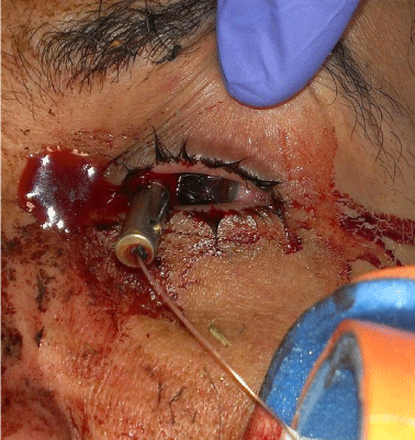
    Figure 1:  Emergency room photograph demonstrating Taser probe injury to
patient’s left eye. Note wire extending from tail-end of probe.
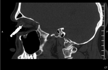
    Figure 2:  Computed tomograph depicting cylindrical, metallic foreign body
inside of a deformed left globe. Note barbed tip at level of posterior sclera.
The patient was taken to the operating room for emergent exploration, removal of ocular foreign body, and open globe repair. Conjunctival peritomy was performed to reveal the entry wound at 10 o’clock, approximately 1mm posterior to limbus. Uveal tissue was noted to be prolapsed around the shaft of the foreign body at the entry wound. The Taser probe was carefully removed while attempting to minimize further damage from the barbed tip of the probe (Figure 3). Exploration of the wound after probe removal revealed a piece of white cylindrical plastic within the globe; this was removed and determined to be the ejector sleeve of the Taser probe. The entry wound was noted to extend superiorly along the limbus to 12 o’clock and medially to the insertion of the medial rectus (Figure 4). Ocular tissue was reposited and the entry wound was closed with nylon suture (Figure 5). An exit wound was not discovered upon globe exploration. One week post-operatively, visual acuity was light perception. The globe was formed with an intraocular pressure of 11 mmHg. Slit lamp examination revealed a shallow chamber with hyphema with no view to the posterior pole. B-scan ultrasonography revealed disorganized intraocular contents. The patient continues to have a significant amount of eye pain and is currently being evaluated for enucleation.
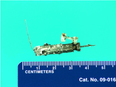
    Figure 3:  Pathology photograph demonstrating Taser probe and plastic
ejector sleeve.
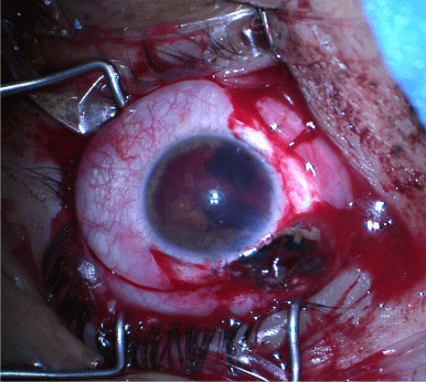
    Figure 4:  Intraoperative photograph after removal of Taser probe. Surgeon’s
view.
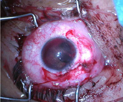
    Figure 5:  Intraoperative photograph after majority of scleral wound repair.
Surgeon’s view.
Discussion
The Taser was invented and developed by National Aeronautics and Space Administration scientist Jack Cover in the 1970s. It is currently produced by Taser International, Inc. and has been used in United States law enforcement since 1998. Tasers and other related Conducted Energy Devices (CEDs) work via administering a high- voltage, low-current electrical charge to disrupt neuromuscular communication, thus incapacitating a suspect. Compressed nitrogen is used to fire two 3 centimeter aluminum probes at approximately 55 meters per second, less than one-fifth the speed of a bullet. Each probe has a 9 meter wire on its tail, used to connect with the power source, and a 9 millimeter long stainless steel barb on the head designed to penetrate clothing and skin. When a closed circuit is completed after both probes have struck the suspect, pulses of electricity with a peak of 1200 volts are sent through the suspect’s body. Reports have shown increasing use of Tasers in law enforcement, from nearly 6,000 U.S. law enforcement agencies deploying Tasers by 2005 to more than 11,000 in 2009 [1]. Taser International’s website reports a more than seven-fold increase in police field-use of Tasers from 2005 to 2011, and currently they estimate more than 900 uses per day worldwide [2]. Texas state law does not recognize the Taser and other CEDs as firearms, and currently there are no restrictions or permits required on the sale or possession for consumers or law enforcement officers alike.
The first report in the literature of Taser related ocular injury was in 2005 [3]. Since the initial report, there have been several other case reports describing a variety of Taser-related eye injuries, mostly due to mechanical trauma. Electrical injury is suggested in two of the case reports: one patient developed an “electrical” cataract attributed to facial Taser injury [4] and the other had an exudative retinal detachment and electroretinogram changes after a Taser penetrated the eyelid of a patient without globe rupture [5]. Penetrating and perforating injuries from Taser probes are the most commonly reported injuries, oftentimes with substantial ocular morbidity [6-9].
Conclusion
With the increasing use of the Taser and other CEDs in law enforcement and elsewhere, we must be aware of the potential for continued eye injuries related to their use. It is important to understand the physical properties of the Taser probe in order to adequately plan for surgical removal. The barbed end may prove to be difficult to remove, especially in the case of posterior globe penetration. We must also be aware of potential damage due to local electric current administered from the Taser probe even in cases without globe penetration.
References
- Police Executive Research Forum. Comparing safety outcomes in police use-of-force cases for law enforcement agencies that have deployed Conducted Energy Devices and a matched comparison group that have not: A quasi-experimental evaluation; Sept 2009. Report submitted to the National Institute of Justice.
- Taser International, Inc. Times Police Have Used TASER CEWs in the Field. 2014.
- Ng W, Chehade M. Taser penetrating ocular injury. Am J Ophthalmol. 2005; 139: 713-715.
- Seth RK, Abedi G, Daccache AJ, Tsai JC. Cataract secondary to electrical shock from a Taser gun. J Cataract Refract Surg. 2007; 33: 1664-1665.
- Sayegh RR, Madsen KA, Adler JD, Johnson MA, Mathews MK. Diffuse retinal injury from a non-penetrating TASER dart. Doc Ophthalmol. 2011; 123: 135-139.
- Teymoorian S, San Filippo AN, Poulose AK, Lyon DB. Perforating globe injury from Taser trauma. Ophthal Plast Reconstr Surg. 2010; 26: 306-308.
- Chen SL, Richard CK, Murthy RC, Lauer AK. Perforating ocular injury by Taser. Clin Experiment Ophthalmol. 2006; 34: 378-380.
- Li JY, Hamill MB. Catastrophic globe disruption as a result of a TASER injury. J Emerg Med. 2013; 44: 65-67.
- Han JS, Chopra A, Carr D. Ophthalmic injuries from a TASER. CJEM. 2009;11: 90-93.