
Case Report
Austin J Clin Ophthalmol. 2015;2(3): 1050.
Incomplete Uveitis – Glaucoma – Hyphema (Ugh) Syndrome in a 36 Year Old Male
Do Nguyen Nguyen1*, Dang Tam Mai2 and Bich Minh Vo Ngoc3
1Doctor, Chief of Phaco Department, HoChiMinh City Eye Hospital, VIetnam
2Doctor, Department of Glaucoma, HoChiMinh City Eye Hospital, Vietnam
3Ophthalmology Intern, Pham Ngoc Thach University of Medicine, Vietnam
*Corresponding author: Do Nguyen Nguyen, Chief of Phaco Department, HoChiMinh City Eye Hospital, Vietnam
Received: April 23, 2015; Accepted: May 28, 2015; Published: June 08, 2015
Abstract
Uveitis – glaucoma - hyphema syndrome (Ellingson syndrome) is a rare condition, associated with anterior or posterior chamber IOLs [1-4]. Recently, most cases of this syndrome have been associated with single-piece acrylic IOLs placed in the sulcus [2]. This condition is caused by the chaffing of the iris with the malpositioned/subluxedIOL [1,3]. The classic triad of UGH syndrome are anterior chamber inflammation with pigment dispersion, increased intraocular pressure and blood in anterior chamber angle weeks to months after cataract surgery [1,3]. The diagnosis is usually difficult, because the clinical manifestations can be incomplete and the patient’s ocular discomfort may not correlate with clinical features. In this paper, we report a case of a patient with posterior chamber IOL who had only two elements of the UGH syndrome.
Abbreviations
IOL: Intraocular Lens; IOP: Intraocular Pressure; OD: Right Eye; OS: Left Eye; PCO: Posterior Capsular Opacification; UGH – Uveitis; Glaucoma; Hyphema; VA: Visual Acuity
Case Report
Patient was 36-year-old Vietnamese male, who presented to Ho Chi Minhcity eye hospital on March, 2015. He had experienced some episodes of transient blurry vision in the right eye for a period of over 12months.
He gave a past ocular history of left eye keratitis 16 years prior to presentation which left a dense corneal scar. He had also undergone phacoemulsification cataract surgery with IOL implantation in his right eye 7 years previously. He had no history of ocular trauma or long term usage of corticoisteroid (either systemic or topical) before the cataract surgery.
The first episode of transient blurrying of vision in the right eye occurred six years after the cataract surgery but was not painful. This was followed by several episodes of blurry vision over the following 12 months. There was no history of ocular trauma postoperatively.
Day 1 pre-op
On the first day when he presented to our hospital, his visual acuity was OD 20/100 and OS hand motion. There was a dense central corneal scar OS (Figure 1B). Examination OD showed no conjunctival injection, poor pupillary reaction, clear cornea, deep anterior chamber, 3+pigmentation in anterior chamber and anterior vitreous and partial dislocation of the sulcus-fixated IOL. The posterior capsule was intact (Figure 1A). Fundus examination revealed the optic nerve with a cup to disc ratio of 0.9 and the thinning of neuroretinal rim (Figure 2). Intraocular pressure was OD 38 mmHg with Goldmann applanation tonometer. Automated Humphrey visual field testing revealed tunnel vision defect in the right eye (Figure 4).
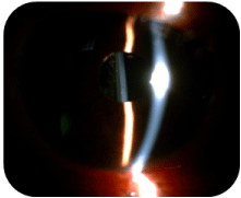
Figure 1A: OD – Deep anterior chamber, 3+ pigmentation in anterior vitreous.
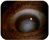
Figure 1B: Dense central corneal scar OS.
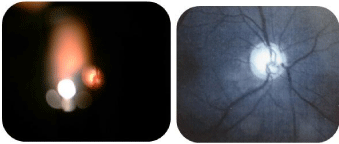
Figure 2: Cup to disc ratio of 0.9 and thinning of neuroretinal rim.
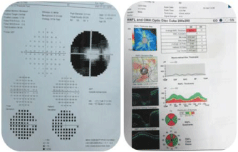
    Figure 4:  Tunnel vision defect and decrease of neuro-retinal fiber thickness
in OCT.
Gonioscopy showed open angle and 3+ pigmentation in the superior, temporal and nasal quarants with heavy pigmentation on the trabecular meshwork inferiorly (Figure 3).
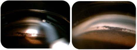
Figure 3: Heavy pigmentation on trabecular meshwork inferiorly.
Topical travoprost/timolol QD, prednisolone acetate 1% QID was used on the right eye and systemic acetazolamide 250mg TID, which reduced the IOP to 14 mmHg.
Day 4 pre-op
The patient reported postural related blurring of vision. The visual acuity was fingers counting at 1 meter OD. Dilation of the eye revealed mobile inferolaterally dislocated one-piece IOL (Figure 5A) which showed contact with the posterior surface of the iris on ultrasound biomicroscopy (Figure 5B). His IOP had, his IOP had, however dropped to 12mmHg. In view of the above findings an IOL exchange was suggested.
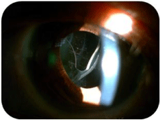
Figure 5A: Dislocation of one-piece IOL after pupil dilate.
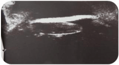
Figure 5B: The contact between IOL and iris in ultrasound biomicroscopsy.
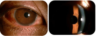
Figure 6: 1 month post-op.
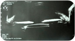
Figure 7: Ultrasound biomicroscopy 1 month post-op – The IOLin position.
Operation day
The patient had the single-piece hydrophobic Acrylic lens in sulcus exchange to a three-piece intraocular lens placed in the bag with no further complications. After the surgery, the patient was treated with topical antibiotic moxifloxacin 0.5% QID and topical prednisolone acetate 1% QID. The IOP-lowering drugs were stopped.
Day 1 post-op
The visual acuity was 20/40 and the IOP was 10mmHg with noncontact tonometer. Examination OD revealed injected conjunctiva, clear cornea, 1+ cell/flare in anterior chamber. The IOL was in position.
Day 4 post-op
The patient reported ocular pain and headache. The visual acuity was 20/50 OD and the IOP was 30 mmHg with Goldmann applanation tonometer. Examination OD revealed injected conjunctiva, clear cornea, no cell/flare or pigmentation in anterior chamber and 1+pigmentation in anterior vitreous. The IOL was in position. Fundus examination showed no change in the optic disc and normal retina.
He continued on topical antibiotic but topical prednisolone acetate 1% was changed to topical nepafenac (NSAIS) QID because of steroid – induced high IOP. Topical brinzolamide 1% TIDwas also used to control the IOP.
1 Week post-op
The patient reported no ocular pain or headache. His visual acuity was 20/25 and the IOP was 10 mmHg. Examination showed clear cornea, clear anterior chamber. He continued with topical NSAID, antibiotic and brinzolamide.
1 Month post-op
The patient had no ocular pain, no episodes of blurry vision. The visual acuity was 20/25 OD and the IOP was 12 mmHg. The gonioscopy showed the significant decrease of pigmentation in trabecular meshwork, especially inferior quadrant (Figures 8A and 8B).
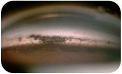
Figure 8A: Gonioscopy before the surgery.

Figure 8B: Gonioscopy 1 month after the surgery.
The patient was maintained on the topical medications with good results.
Discussion
The single-piece hydrophobic acrylic lens has square edges that have been shown to reduce the incidence of PCO if placed in the capsular bag, but when placed in the ciliary sulcus, the square edges of the lens and the haptic may make contact with the posterior iris and ciliary sulcus uveal tissue, especially with the dilation and constriction of the iris. These may cause pigment dispersion. The interesting characteristics seen in this case is the pigment dispersion in the anterior chamber, vitreous cavity and trabecular meshwork. Associated with this finding is usually a rise in IOP, indicating pigment dispersion syndrome secondary to the IOL. This syndrome is thought to occur when contact exists between the IOL and the posterior iris, known as “iris chaffing”, which causes excess liberation of pigment from the iris epithelium with subsequent obstruction of the trabecular meshwork outflow pathway. With time, this leads to pseudophakic glaucoma; a very serious complication of cataract surgery.
Conclusion
This case illustrates the postoperative complications of uveitis with pigment dispersion and glaucoma following single-piece IOL placed in the ciliary sulcus. Single-piece IOL should not be placed in the ciliary sulcus, but should be implanted only in the capsular bag as recommended by the manufacturer in order to avoid complications later.
References
- American Academy of Ophthalmology. "Lens and cataract: section 11, basic and clinical science course". San Francisco, USA. 2011.
- Chang DF, Masket S, Miller KM, Braga-Mele R, Little BC, Mamalis N, et al. Complications of sulcus placement of single-piece acrylic intraocular lenses: recommendations for backup IOL implantation following posterior capsule rupture. Journal of Cataract & Refractive Surgery. 2009; 35: 1445-1458.
- Stamper, Robert L, Lieberman MF, Michael V. Drake. Becker-Shaffer's diagnosis and therapy of the glaucomas. Elsevier Health Sciences. 2009.
- Van Liefferinge T, Van Oye R, Kestelyn P. Uveitis-glaucoma-hyphaema: a late complication of posterior chamber lenses. Bull SocBelgeOphthalmol. 1994; 252: 61–65.