
Research Article
Austin J Clin Ophthalmol. 2015; 2(5): 1058.
Correlation between Diabetic Macular Edema and Peripheral Retinal Ischemia in Saudi Arabia
Al Shammari AS¹ and El-Agamy AF²*
¹Department of Optometry and Vision Sciences, College of Applied Medical Sciences, King Saud University, Saudi Arabia
²Department of Optometry and Vision Sciences, College of Applied Medical Sciences, King Saud University, Saudi Arabia and Faculty of Medicine, Mansoura University, Egypt
*Corresponding author: El-Agamy AF, Department of Optometry and Vision Sciences, College of Applied Medical Sciences (female section), King Saud University, Building 11, Third Floor, Office 223, Riyadh, Saudi Arabia
Received: August 19, 2015; Accepted: October 01, 2015; Published: October 08, 2015
Abstract
Evaluation of the correlation between Peripheral Retinal Ischemia (PRI) and Diabetic Macular Edema (DME)in Saudi patients using Ultra-Wide Field Fluorescein Angiography (UWFA) and Spectral domain Optical Coherence Tomography (OCT) imaging techniques.
Subjects and Methods: A prospective non- randomized cross sectional study of 90 eyes of 58 Saudi Diabetic Retinopathy (DR) patients (36 Males and 22 Females aged from 22-80 years old) were examined using diagnostic UWFA (Optos 200Tx imaging system), in the late phase of Fluorescein Angiography (FA) to assess the presence of PRI. Based on OCT and early/ mid phases of FA using Topcon camera, patients were classified as either having DME or not. Chi-square test was used to assess the correlation between DME and PRI. The level of statistical significance was set at (P= 0.05).
Results: This study included 90 eyes from 58 DR patients (36 Males and 22 Females) who underwent diagnostic UWFA. DME was detected in 69 eyes (76.7%) of those eyes. Eyes diagnosed with DME were divided into four types: diffuse 36 eyes (40.0%); focal 23 eyes (25.6%); combined 6 eyes (6.7%) and cystoids, 4 eyes (4.4%). Out of these 90 eyes, 63 (70.0%) demonstrated PRI.
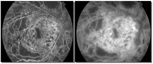
Figure 1: Early/Mid phase of diffuse leakage on FA by Topcon camera.
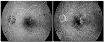
Figure 2: Early/Mid phase of focal leakage on FA by Topcon camera.
PRI was detected in 32 eyes with diffuse DME (64.0%), 12 eyes with focal DME (24.0%), 4 eyes with combined DME (8.0%), and 2 eyes with cystoids DME (4.0%).
In this study, there was a statistically significant relationship between the presence of PRI and DME in diabetic eyes, especially the diffuse type (P value= 0.013). There was median correlation (0.394) between PRI and DME types.
Conclusion: There is a significant correlation between DME and PRI. UWFA is a useful tool for detecting PRI which may have direct implications in the diagnosis, follow-up and treatment such as targeted peripheral photocoagulation.
Keywords: Peripheral retinal ischemia; Diabetic macular edema; Ultrawide field fluorescein angiography; Optical coherence tomography; Diabetic retinopathy
Introduction
Diabetic Macular Edema (DME) is the most common cause of visual loss in patients with Non-Proliferative Diabetic Retinopathy (NPDR) [1]. According to the Wisconsin Epidemiologic Study of Diabetic Retinopathy (DR), the prevalence of DME after 15 years of diagnosed diabetes is around 20% in patients with type 1 diabetes, 25% in patients with insulin-dependent type 2 diabetes, and 14% in patients with non-insulin dependent type 2 diabetes [2].
The conventional Fundus camera provides good photographs of optic nerve and posterior pole but with a limited view of the retinal periphery. The invention of Ultra-Wide Field Fluorescein Angiography (UWFA) in 2000 overcomes this limitation. It is capable to image 200 internal degrees field of view. An ellipsoid mirror is used to image the retinal periphery. A red (633 nm) and green laser (523 nm) was utilized to scan the retina and obtain the images. In addition, high-resolution Fluorescein Angiography (FA) images of the retinal periphery obtained by Optos are clinically useful in eyes with retinal vascular disease [3].
Optical Coherence Tomography (OCT) is a non-invasive and noncontact diagnostic method for detection of various retinal diseases. It uses infrared light reflections to obtain reliable, reproducible, and objective cross sectional images of the retinal structures and the vitreoretinal interface and allows quantitative measurements of retinal thickness [2].
The aim of this cross sectional study is to assess the correlation between Peripheral Retinal Ischemia (PRI) and DME in Saudi DR Patients using UWFA and Spectral domain OCT imaging techniques.
Subjects and Methods
This study was a prospective non-randomized cross sectional study. 90 eyes of 58 Saudi DR patients (36 Males and 22 Females aged from 22-80 years old at King Khaled Eye Specialist Hospital (KKESH) were enrolled in the study (from January to June 2013). They were examined using diagnostic UWFA (Optos 200Tx imaging system) in the late phase of FA to assess the presence of PRI. Based on OCT and early / mid phases of FA using Topcon camera, patients were classified as either having DME or not.
Different types of DME were differentiated according to the pattern of leakage related to the vascular abnormalities within or adjacent to the Fluorescein dye leakage areas. Focal macular edema was defined by the presence of localized areas of retinal thickening associated with focal leakage of micro aneurysm/s. Diffuse macular edema was identified by a more generalized form of edema, visualized as widespread macular leakage and pooling of dye in cystic spaces. CME was diagnosed by accumulation of dye in the perifoveal region in a petallloid pattern [2]. DME in OCT scans was discriminated by diffuse thickening of the neurosensory retinal and loss of the foveal depression, cystic retinal changes, which manifested as areas of low intraretinal reflectivity; and serous retinal detachment, alone or combined [2].
Inclusion criteria of the DR patients were more than 18 years old and able to understand instructions.
Exclusion criteria are patients with a history of focal, pan-retinal laser photocoagulation or retinal surgeries. In addition, patients with history of vascular occlusions, uveitis, sickle cell retinopathy, retinal scars, intraocular tumors, significant media opacity, vitreous hemorrhage and ptosis were excluded.
The research followed the tenets of the Declaration of Helsinki that informed consent was obtained from the subjects after explanation of the nature and possible consequences of the study. The research was approved by the appropriate Institutional Review Board (IRB).
The research followed the tenets of the Declaration of Helsinki that informed consent was obtained from the subjects after explanation of the nature and possible consequences of the study. The research was approved by the appropriate Institutional Review Board (IRB).
The selected patients were injected by 5 cc of sodium Fluorescein dye (10%). The images were captured first by Topcon camera followed immediately by UWFA. It provides high resolution images of about 82.5 % of the total retinal surface area in a single capture [4]. All captured images were compressed in to high quality jpeg files, and transferred to Adobe soft-ware for review [5]. V2 vantage Pro contains a set of applications that monitor and manage the quality of images. Each eye was evaluated for presence or absence of PRI in Optos images.
OCT images were analyzed to reveal the presence or absence of DME in those eyes. Macular thickness and volume were measured by Heidelberg Eye Explorer Soft Ware.
Chi-square test depending on the frequency values was used to assess the correlation between DME and PRI. The level of statistical significance was set at (P= 0.05). Frequency and percentage were calculated for categorical variables like: DME (present or absent), type of DME and PRI. Finally, SPSS version 16 system was used to do all statistical parts in this study.
Results
A total of 126 eyes of 63 diabetic patients were examined by UWFA at photography department of KKESH in Kingdom of Saudi Arabia (K.S.A). Thirty six eyes were excluded according to the exclusion criteria. This study included 90 eyes 200Tx imaging from 58 DR patients (36 Males and 22 Females) who underwent diagnostic UWFA.
DME was detected in 69 eyes (76.7%) of those eyes (Table1). Eyes diagnosed with DME were divided into four types: diffuse (Figure 1) 36 eyes (40.0%); focal (Figure 2) 23 eyes (25.6%); combined (Figure 3) 6 eyes (6.7%) and cystoids 4 eyes (4.4%) (Graph 1).
    Diabetic Macular Edema (DME)
    Frequency
    Percent


    Present
    69
    76.7


    Absent
    21
    23.3


    Total
    90
    100.0
Table 1: DME was detected in 69 eyes of the total eyes.
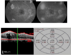
Figure 3: Combined pattern on FA and OCT.
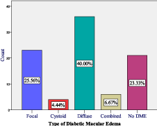
    Graph 1:   Bar chart illustrates types of detected DME. 36 Diffuse, 23 Focal, 6
Combined, and 4 CME. Diffuse DME type was of the highest value compared
to other types of DME.
Out of the 90 eyes, 63 eyes (70.0%) demonstrated PRI (Table 2) and (Figure 4). PRI was detected in 32 eyes with diffuse DME (64.0%), 12 eyes with focal DME (24.0%), 4 eyes with combined DME (8.0%), and 2 eyes with cystoids DME (4.0%).
    Peripheral retinal Ischemia
    Frequency
    Percent


    Present
    63
    70.0


    Absent
    27
    30.0


    Total
    90
    100.0
Table 2:  Frequency table shows the number and percentage of eyes with and
without PRI.
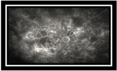
    Figure 4:  Ischemic areas detected as areas of hypo-fluorescence in the
periphery by Optos camera.
In this study, there was a statistically significant relationship between the presence of PRI and DME in diabetic eyes, especially the diffuse type of DME (P value= 0.013). There was median correlation (0.394) between PRI and DME types (Table 3).
    Peripheral Retinal Ischemia (PRI)
    Type of DME
    Total
      69(100.0%)
    Pearson Chi-Square
    Correlation
    Significance


    Diffuse
      36(52.2%)
    Focal
      23(33.3%)
    Combined
      6(8.7%)
    Cystoid
      4(5.8%)


    Present
    32(88.9%)
    12(52.2%)
    4(66.7%)
    2(50.0%)
    50(72.5%)
    10.725
    0.394
    0.013*


    Absent
    4(11.1%)
    11(47.8%)
    2(33.3%)
    2(50.0%)
    19(27.5%)
Table 3: Shows the relationship between PRI and DME types with median correlation between them which equals to 0.394.
Discussion
In DR, Ischemic changes lead to the production of Vascular Endothelial Growth Factor (VEGF) [6], which can cause the breakdown of blood-retinal barriers, and increased retinal vessel permeability producing DME. The successful effect of anti-VEGF therapy in the management of DME may support the idea that retinal ischemia and DME are correlated [7]. But assessment of PRI using conventional retinal imaging modalities is challenging. UWFA has become a valuable tool for detection of PRI [8-10]. Since first described by Friberg and Forrester in 2004 [11], UWFA has proven to have favorable sensitivity and specificity compared to other traditional methods including 7 standard fields (7SF) [12,13]. Recent comprehensive study showed the good ability of UWFA imaging system in detection of diabetic pathology [10]. The same study found that 7SF has failed to detect either ischemia or neovascularization in 10% of eyes. Visualization of up to 200 degrees or more of the peripheral retina captured in a single frame can be obtained using UWFA imaging system [10].
In this study, PRI was detected in 70%of the examined eyes. This was almost equal to what conducted by Mackenzie PJ when they examined the sensitivity and specificity of the Optos Opto map for detection of peripheral retinal lesion in (2007) [13].
In our study, DME types were detected by both imaging systems (FA and OCT). This is because there is a discrepancy between OCT and FA outcomes that was mentioned in Ozdek et al. [14]. In their study, diabetic CME was detected with Stratus OCT in 30 eyes (15.4%) but not confirmed by FA in 63.3% of these cases. 3D-OCT is more sensitive and reproducible than FA for the detection of CME type [13]. In addition, OCT is superior in evaluating foveal changes that are not evident in diabetic eyes angiographically [15]. In contrast; OCT cannot detect the abnormal vessels and abnormal blood flow which can be imaged by FA only [16].
In our study, FA and OCT showed 76.7% of the examined eyes with DME and 23.3% of eyes showed no evidence of DME. There is a variation in time between capturing Fluorescein images and OCT images. This may affect the Fluorescein leakage patterns especially in the diffuse type. We recommend a one machine combining both FA and OCT in order to help in a specific definition of pathophysiology (FA) and morphologic (OCT) retinal changes related to DME. This agreed with the recommendations of Bolz et al. [16].
Witmer et al., compared Optos Optomap with Heidelberg Spectralis OCT. They are UWF imaging devices. Optos Optomap was found to be effective in imaging the temporal and nasal retina compared to OCT which was superior in evaluating the superior and inferior retina to farther point [8].
The limitations in our study were the duration was only 3 months, and it was a random study including DR eyes existed in outpatient clinics in KKESH.
Conclusion
There is a significant correlation between DME and PRI. UWFA is a useful tool for detecting PRI which may have direct implications in the diagnosis, follow-up and treatment such as targeted peripheral photocoagulation.
References
- Duh EJ. Diabetic retinopathy. Totowa, New Jersey; Humana Press. 2008.
- Lobo C, Pires I, Cunha-Vaz J. Diabetic Macular Edema. In Optical Coherence Tomography. Springer, Berlin, Heidelberg. 2012; 1-21.
- Witmer MT, Kiss S. The Clinical Utility of Ultra-Wide-Field Imaging: a look at four widely available methods of imaging the peripheral retina that are making photography more clinically practical. New York City; 2012.
- Optos. (April 2013).
- Optos handbook. Introductory Handbook. United Kingdom. OptosPlccompany.By: Optosplc, Queensferry House, Carnegie Campus, Dunfermline, Scotland, KY11 8GR, United Kingdom.
- Aiello LP, Avery RL, Arrigg PG, Keyt BA, Jampel HD, Shah ST, et al. Vascular endothelial growth factor in ocular fluid of patients with diabetic retinopathy and other retinal disorders. N Engl J Med. 1994; 331: 1480-1487.
- Haritoglou C, Kook D, Neubauer A, Wolf A, Priglinger S, Strauss R, et al. Intravitreal bevacizumab (Avastin) therapy for persistent diffuse diabetic macular edema. Retina. 2006; 26: 999-1005.
- Witmer MT, Kiss S. Wide-field imaging of the retina. Survey of ophthalmology. 2013; 58: 143-154.
- Wessel MM, Aaker GD, Parlitsis G, Cho M, D'Amico DJ, Kiss S. Ultra—Wide-Field Angiography improves the detection and classification of Diabetic Retinopathy. Retina. 2012; 32: 785-791.
- Wessel MM, Nair N, Aaker GD, Ehrlich JR, D'Amico DJ, Kiss S. Peripheral retinal ischemia, as evaluated by ultra-wide field Fluorescein angiography, is associated with diabetic macular odema. British Journal of Ophthalmology. 2012; 96: 694-698.
- Friberg TR, Gupta A, Yu J, Huang L, Suner I, Puliafito CA, et al. Ultra wide angle fluorescein angiographic imaging: a comparison to conventional digital acquisition systems. Ophthalmic surgery lasers imaging: the official journal of the International Society for Imaging in the Eye. 2008; 39: 304-311.
- Mackenzie PJ, Russell M, Ma PE, Isbister CM, AL Maberley DA. Sensitivity and specificity of the OptosOptomap for detecting peripheral retinal lesions. Retina. 2007; 27: 1119-1124.
- Ouyang Y, Keane PA, Sadda SR, Walsh AC. Detection of cystoids macular edema with three-dimensional optical coherence tomography versus Fluorescein angiography. Investigative ophthalmology & visual science. 2010; 51: 5213-5218.
- Ozdek SC, Erdinç MA, Gürelik G, Aydin B, Bahçeci U, Hasanreisoğlu B. Optical coherence tomographic assessment of diabetic macular edema: comparison with Fluorescein angiographic and clinical findings. Ophthalmologica. 2005; 219: 86-92.
- Johnson MW. Tractional cystoids macular edema: a subtle variant of the vitreo macular traction syndrome. Am J Ophthalmol. 2005; 140: 184 -192.
- Bolz M, Ritter M, Schneider M, Simader C, Scholda C, Schmidt-Erfurth U. A systematic correlation of angiography and high-resolution optical coherence tomography in diabetic macular edema. Ophthalmology. 2009; 116: 66-72.