
Research Article
Austin J Clin Ophthalmol. 2016; 3(2): 1070.
Correlation between Anthropomorphic Measurements and Ocular Parameters among Adult Saudi Females
Fahmy RM1,2*
¹Department of Ophthalmology, Faculty of Medicine, Cairo University, Egypt
²Department of Optometry, College of Applied Medical Sciences, King Saud University, Saudi Arabia
*Corresponding author: Fahmy RM, Department of Optometry, King Saud University, Saudi Arabia
Received: May 23, 2016; Accepted: July 21, 2016; Published: July 28, 2016
Abstract
Purpose: To evaluate the correlation of anthropomorphic measurements (height, weight and body mass index) with ocular parameters among adult Saudi females.
Methods: The study included 155 females (155 eyes), age ranging from 18 to 27 years. Pentacam Scheimpflug images were used to measure Anterior Chamber Depth (ACD) and central corneal thickness. IOL Master was used to measure Axial Length (AL). An autorefracto/kerato/tonometer was used to measure the Intraocular Pressure (IOP) and Refraction/Spherical Equivalent (SE). The anthropomorphic measurements body height and weight have been measured using a wall-mounted metric ruler and digital floor scale, respectively. Body Mass Index (BMI) was calculated as weight divided by square of height.
Results: Body height was significantly associated with higher body weight (r = 0.398, p = <0.001). Height correlated positively with axial length (r = 0.203, p = 0.011). Central corneal thickness, ACD, SE and IOP were not significantly associated with body height. Body weight was significantly associated with higher BMI (r = 0.941, p = <0.001). Body mass index and body weight were not significantly associated with all ocular parameters. Significant negative correlation was found between age and ACD (r = -0.307, p = <0.001). Also, significant positive correlation was found between AL and ACD (r = 0.444, p = <0.001), as well as between CCT and IOP (r = 0.357, p = <0.001). Significant negative correlation was found between AL and SE (r = -0.608, p = <0.001), as well as between ACD and SE (r = 0.330, p = <0.001).
Conclusion: The results indicate that there is a significant correlation between height and AL in this adult population. Also, it confirms a negative correlation between AL and SE, and positive correlation between AL and ACD, as well as between CCT and IOP.
Keywords: Axial length; Anterior chamber depth; Body height; Body mass index; Central corneal thickness; Intraocular pressure; Spherical equivalent
Introduction
The field of anthropomorphic encompasses a variety of human body measurements, such as height, weight, and Body Mass Index (BMI), circumferences, skin fold thicknesses, lengths and breadths. Body measurement data in adults are used to evaluate health and dietary status, disease risk and body composition changes that occur over the adult lifetime [1].
Average height is frequently characteristic within the group when populations share genetic background and environmental factors. There are exceptional height variation within populations such as dwarfism or gigantism, which are medical conditions caused by specific genes or endocrine abnormalities [2]. Body mass index is a measure of weight adjusted for height, simple to calculate as weight in kilogram divided by the square of height in meters (kg/m²). Body mass index levels correlate with body fat and with future health risks [3].
Anthropomorphic measurement of height, weight or calculated BMI can be associated with ocular parameter. Ocular parameters are used to diagnose diseases and diseases development.
Study performed in the rural population of Central India found that body height and size of the eyes were associated with each other, where taller subjects had larger eyes with flatter corneas. An increase in body height per 10 cm was associated with an increase in anterior chamber depth by 1% and an increase in vitreous cavity length by 1%. Subjects with a higher BMI had shorter eyes, flatter and thicker corneas. Taller subjects and subjects with a higher BMI were more hyperopic [4].
In open-angle glaucoma, the anterior chamber angle is opened and the ACD is normal as assessed by slit-lamp biomicroscopy and gonioscopy. In angle-closure glaucoma, the chamber angle is either occluded by ≥ 15 degrees or the peripheral ACD is ≤ 25% of corneal thickness. The Beijing Eye Study that aimed to assess differences in anthropomorphic measures between POAG and PACG, they found that PACG was significantly associated with shorter body height, age, hyperopic refractive error, female gender and a shallower anterior chamber. However, it did not vary significantly in terms of body weight, BMI and optic disc area. The only parameter to retain a significant difference between the two glaucoma groups was ACD. Age, gender, body height, refractive error and level of education were no longer significantly associated with either of the two glaucoma groups [5].
In our study, we were evaluating the relationship of anthropomorphic parameters including the height, weight and BMI with ocular parameters among adult Saudi females. The results may be helpful in inclusion of body parameters in the list of diagnostic variables and risk factors of some ocular diseases such as angleclosure glaucoma.
Subjects and Methods
This study involves 155 healthy (155 eyes) Saudi Arabian female subjects. It was conducted from the first of October 2015 until the 10th of December 2015, at King Saud University, College of Applied Medical Sciences (female campus). The mean age was 20.63 ± 1.529 years old and the range was between 18 to 27 years old. Patients with cataract, glaucoma, anterior segment inflammation or any systemic diseases with ocular complications were excluded. After full general health and ocular history taking, a body height (in meter) and weight (in kilogram) have been measured using a wall-mounted metric ruler and digital floor scale, respectively. Body mass index was calculated as weight divided by the square of the height (kilograms per square meter).
Ocular parameters measurements were performed using: Auto Kerato/Refracto/Tonometer TRK-1P from Topcon to measure the refraction (SE) and IOP.
IOL Master to measure the axial length.
Oculus Pentacam HR to measure the central corneal thickness and anterior chamber depth.
Statistical Analysis
Statistical analysis was performed by using a commercially available statistical software package (SPSS for Windows, version 22.0). Measurable data of the study was presented as mean ± Standard Deviation (SD). The association between clinical measurements was analyzed by Pearson’s sample correlation coefficient (r) test. All p-values were two-sided and were considered statistically significant when the values were less than 0.05.
Results
Descriptive analysis
The mean age of subjects was 20.63 ± 1.529 years. The mean body height was 1.59 ± 0.057 m (range: 1.45 to 1.76 m), and the mean body weight was 59.83 ± 13.307 kg (range: 40 to 111.9 kg), resulting in a mean BMI of 23.97 ± 4.859 kg/m2 (range: 16.023 to 42.638 kg/m²). The mean Spherical Equivalent (SE) of refractive error was -1.076 ± 1.862 diopters (range: -7.50 to +7.75 diopters) (Table 1).
    Parameters
    Mean ± SD
    Range


    Age (years)
    20.63 ± 1.529
    18 - 27


    Anthropomorphic    measurements
      Height (m)
      Weight (kg)
      BMI (kg/m2)

      1.59 ± 0.057
      59.83 ± 13.307
      23.97 ± 4.859

      1.45 – 1.76
      40 – 111.9
      16.023 – 42.638


    Ocular measurements
      SE (D)
      IOP (mmHg)
      AL (mm)
      ACD (mm)
      CCT (µm)

      -1.076 ± 1.862
      18.87 ± 2.763
      23.75 ± 1.014
      3.51 ± 0.325
      555.54 ± 31.714

      -7.50 – 7.75
      13 – 24
      21.42 – 26.31
      2.46 – 4.35
      471 – 637
Table 1: Data of subjects.
Correlation between age and ocular parameters
A moderate but significant negative correlation was found between age and ACD (r = -0.307, p = <0.001) (Figure 1). Axial length, SE, CCT and IOP were not significantly associated with age (Table 2).
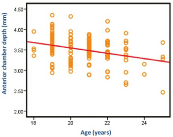
Figure 1: Scatter plot between ACD and Age.
    Parameters
    SE
    AL
    ACD
    CCT
    IOP


    Age correlation coefficient
      p-value
    0.004
      0.959
    0.014
      0.863
    -0.307
      <0.001*
    0.107
      0.187
    -0.148
      0.066


    *p-value statistically significant
Table 2: Association (bivariate analysis) between age and ocular parameters.
Body height with clinical parameters results
Moderate significant positive correlation was found between body height and body weight (r = 0.398, p = <0.001). There was a positive weak correlation between body height and AL (r = 0.203, p = 0.011) (Figure 2). Central corneal thickness, IOP, ACD and SE were not significantly associated with body height (Table 3).
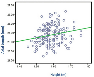
Figure 2: Scatter plot between AL and height measurements.
    Parameters
    Correlation coefficient
    p-value


    Body weight
      Body mass index
      Spherical equivalent
      Intraocular pressure
      Axial length
      Anterior chamber depth
      Central corneal thickness
    0.398
      0.070
      0.024
      -0.009
      0.203
      -0.020
      0.068
    <0.001*
      0.387
      0.685
      0.911
      0.011*
      0.806
      0.398


    *p-value    statistically significant
Table 3:  Association (bivariate analysis) between body height (measured in m)
and clinical parameters.
Body weight and BMI with clinical parameters results
Strong significant positive correlation was found between body weight and BMI (r = 0.941, p = <0.001). Body mass index and body weight were not significantly associated with all ocular parameters (Table 4 and Table 5).
    Parameters
    Correlation coefficient
    p-value


    Body height
      Body mass index
      Spherical equivalent
      Intraocular pressure
      Axial length
      Anterior chamber depth
      Central corneal thickness
    0.398
      0.941
      0.070
      0.109
      0.076
      -0.037
      0.077
    <0.001*
      <0.001*
      0.387
      0.177
      0.347
      0.649
      0.339


      *p-value statistically significant
Table 4:  Association (bivariate analysis) between body weight (measured in kg)
and clinical parameters.
    Parameters
    Correlation coefficient
    p-value


    Body height
      Body weight
      SE
      Intraocular pressure
      Axial length
      ACD
      CCC
    0.070
      0.941
      0.074
      0.121
      0.006
      -0.033
      0.062
    0.387
      <0.001*
      0.357
      0.133
      0.938
      0.684
      0.440


      *p-value statistically    significant
Table 5:  Association (bivariate analysis) between BMI (measured in kg/m2) and
clinical parameters.
Correlation between ocular parameters
A moderate but significant positive correlation was found between AL and ACD (r = 0.444, p = <0.001) (Figure 3), as well as between CCT and IOP (r = 0.357, p = <0.001) (Figure 4). A moderate but significant negative correlation was found between AL and SE (r = -0.608, p = <0.001) (Figure 5), as well as between ACD and SE (r = -0.330, p = <0.001) (Figure 6) (Table 6).
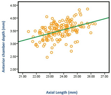
Figure 3: Scatter plot between ACD and AL measurements.
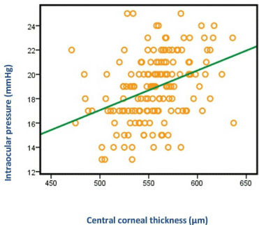
Figure 4: Scatter plot between IOP and CCT measurements.
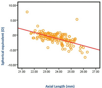
Figure 5: Scatter plot between SE and AL measurements.
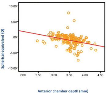
Figure 6: Scatter plot between SE and ACD measurements.
    Parameters
    SE
    AL
    ACD
    CCT
    IOP


    AL
    correlation    coefficient
    -0.608
    1
    0.444
    0.150
    -0.026


    p-value

    <0.001*
    <0.001*
    0.062
    0.748


    ACD
    correlation    coefficient
    -0.330
    0.444
    1
    0.074
    0.075


    p-value

    <0.001*
    <0.001*
    0.363
    0.355


    CCT
    correlation    coefficient
    -0.004
    0.150
    0.074
    1
    0.375


    p-value

    0.964
    0.062
    0.363
    <0.001'


    IOP
    correlation    coefficient
    0.026
    -0.026
    0.075
    0,375
    1


    p-value
    0.752
    0.748
    0.355
    <0,001*


    *p-value statistically significant
Table 6: Association (bivariate analysis) between ocular parameters.
Discussion
In this study, there was a positive correlation between body height and body weight (r = 0.398, p = <0.001). Also, there was a positive correlation between body height and AL (r = 0.203, p = 0.011). Central corneal thickness, IOP, ACD and SE were not significantly associated with body height. Body weight was significantly associated with higher BMI (r = 0.941, p = <0.001). Body mass index and body weight were not significantly associated with all ocular parameters.
Age correlated negatively with ACD (r = -0.307, p = <0.001). Positive correlation was found between AL and ACD (r = 0.444, p = <0.001), as well as between CCT and IOP (r = 0.357, p = <0.001). A significant negative correlation was found between AL and SE (r = -0.608, p = <0.001), as well as between ACD and SE (r = 0.330, p = <0.001).
In Singapore Chinese adults aged 40 to 81 years, Wong et al. [3] found that adult height was related to ocular dimensions, but does not appear to influence refraction. Taller persons are more likely to has longer AL (r = 0.333, p = <0.001), deeper ACDs (r = 0.311, p = <0.001), thinner lenses (r = -0.242, p = <0.001) and flatter corneas (r = 0.301, p = <0.001). This study is consistent with the current study except for the correlation between height and ACD. On the other hand, they found that obese adults were mildly more hyperopic (r = 0.100, p<0.01), and this does not correspond to the current study. Similarly, taller Singapore Chinese children aged 7 to 9 years had longer ALs, thinner lenses, deeper ACDs, flatter corneas and more myopic refraction. However, obese children had refractions tend toward hyperopia [6]. The discrepancies between the results of the two studies could be due to different sample sizes, different ethnicity, age range and refractive error measurement techniques (Table 7).
Author
Population
N
Age
Method
Ocular parameters
Wong et al. [3]
Singapore
(Adults)951
40-81
A-scan Ultrasound
AL*
ACD*
SE
CR*Saw et al. [6]
Singapore
(Children)1449
7-9
A-scan Ultrasound
AL*
ACD*
SE*
CR*Eysteinsson et al. [8]
Iceland
(Adults)832
55-100
A-scan Ultrasound
AL*
ACD
SE
CR*Ojainu et al. [7]
Australia
(Children)1765
6-13
IOL Master
AL*
ACD
SE
CR*Xu et al. [9]
China
(Adults)3191
40
Van Herick
ACD*
Nangia et al. [4]
Central
India
(Adults)4711
30-74
A-scan Ultrasound
AL*
ACD*
SE
CR*
CCT
IOPXu et al. [10]
China
(Adults)3251
45-89
-
ACD*
CCT* 12
SE
IOPGunes et al. [11]
Turkey
(Adults)68
27-69
LenStar
AL
ACD
CCT
IOP*Current study
Saudi
Arabia
(Adults)155
18-27
IOL Master
AL*
ACD
CCT
SE
IOP*Significant correlation between ocular parameter and height
significant correlation between ocular parameter and BMI
Table 7: Different sample sizes, different ethnicity, age range and refractive error measurement techniques.
Ojaimi et al. [7] found that height was strongly related to AL (r = 0.252, p = <0.001) and Corneal Radius (CR) (r = 0.205, p = <0.001). However, there was no significant association between refraction and any of the measured anthropomorphic parameters in Australian children. Moreover, Eysteinsson et al. [8] found that height correlated positively with AL (p<0.01) and CR (p = <0.001). They found a significant negative correlation between AL and SE (r = -0.595, p<0.001), and between age and ACD (p = <0.001). Also, weight was unrelated to all ocular parameters. These findings are, to a great extent, consistent with the results of the current study.
In the rural population of Central India aged 30 to 74, body height correlated positively with AL (p = 0.03) and ACD (p = 0.006), and negatively with CR (p = <0.001). Central corneal thickness (p = 0.44), IOP (p = 0.87) and SE (p = 0.28) were not significantly associated with body height. The BMI, when compared with body height, had a markedly lower influence on all ocular parameters [4]. This study is also consistent with the current study except for the correlation between height and ACD.
Xu et al. [9] suggested that taller Chinese adults had deeper peripheral ACD (p = <0.001) using van Herick’s method. Weight and BMI were not significantly associated with peripheral ACD (p = 0.97) (p = 0.82), respectively. It confirms a recent report from the Beijing Eye Study, in which body height was significantly associated with ACD (p = <0.001) and CCT (p = <0.001). However, it was not associated with IOP (p = 0.99) and SE (p = 0.40) [10]. These studies are consistent with the current study except for the correlation between height and ACD.
There was a positive correlation between BMI and IOP (r = 0.404, p < 0.001). ACD was negatively correlated with BMI. However, BMI was not associated with AL and CCT [11]. This study is inconsistent with the current study due to difference in mean BMI (30.60 kg/m2 vs. 23.97 kg/m2).
The limitation of the current study is that the studied subjects were females only, so it cannot provide information about the effect of anthropomorphic measurements on ocular parameters of males. In contrast to some previous population based studies, the strength of the current study is the inclusion of relatively young subjects with an average age of 20.63 ± 1.5 years modifying the ACD.
Conclusion and Recommendations
In conclusion, AL was significantly longer in taller subjects. Central corneal thickness, IOP, ACD and SE were not significantly associated with body height. Weight and BMI were not associated with all ocular parameters. All previous studies found a significant correlation between anthropomorphic measurements and ocular parameters due to their large sample size. These previous results help the optometrists in screening purpose. On the other hand, current study was evaluating small sample size helping in the assessment of risk factors, diagnosis and treatment of some ocular diseases. A further longitudinal study over large sample Saudi population is recommended to be performed.
References
- Fryar C, Gu Q, Ogden CL. Anthropometric Reference Data for Children and Adults : United States, 2007–2010. National Center for Health Statistics. Vital Health Stat. 2012.
- Ganong WF. Review of Medical Physiology. 12th Edn. California: Lange Medical. 2001.
- Wong TY. Foster PJ, Johnson GJ, Klein BE, Seah SK. The relationship between ocular dimensions and refraction with adult stature: The Tanjong Pagar survey. Investigative Ophthalmology and Visual Science. 2001; 42: 1237-1242.
- Nangia V, Jonas JB, Matin A, Kulkarni M, Sinha A, Gupta R. Body height and ocular dimensions in the adult population in rural central India. The Central India Eye and Medical Study. Graefe’s Archive for Clinical and Experimental Ophthalmology. 2010; 248: 1657-1666.
- Xu L, Li J, Wang Y, Jonas JB. Anthropomorphic differences between angle-closure and open-angle glaucoma: The Beijing Eye Study. Acta Ophthalmologica Scandinavica, 2007; 85: 914-915.
- Saw SM, Chua WH, Hong CY, Wu HM, Chia KS, Stone RA, et al. Height and its relationship to refraction and biometry parameters in Singapore Chinese children. Investigative Ophthalmology and Visual Science. 2002; 43: 1408- 1413.
- Ojaimi E, Morgan IG, Robaei D, Rose KA, Smith W, Rochtchina E, et al. Effect of stature and other anthropometric parameters on eye size and refraction in a population-based study of Australian children. Investigative Ophthalmology and Visual Science. 2005; 46: 4424-4429.
- Eysteinsson T, Jonasson F, Arnarsson A, Sasaki H, Sasaki K, et al. Relationships between ocular dimensions and adult stature among participants in the Reykjavik Eye Study. Acta Ophthalmologica Scandinavica. 2005; 83: 734-738.
- Xu L, Li JJ, Xia CR, Wang YX, Jonas JB. Anterior chamber depth correlated with anthropomorphic measurements: the Beijing Eye Study. Eye (London, England). 2009; 23: 632-634.
- Xu L, Wang YX, Zhang HT, Jonas JB. Anthropomorphic measurements and general and ocular parameters in adult Chinese: The Beijing Eye Study. Acta Ophthalmologica. 2011; 89: 442-447.
- Gunes A, Uzun F, Karaca EE, Kalaycı M. Evaluation of Anterior Segment Parameters in Obesity. Korean J Ophthalmol. 2015; 29: 220-225.