Case Presentation
Austin J Clin Ophthalmol. 2016; 3(3): 1071.
Lemierre Syndrome Presenting with Unilateral Orbital and Cavernous Sinus Thrombosis
Zhang R¹, Chon BH¹* and Collins AC²
¹Department of Ophthalmology, University Hospitals Case Medical Center, USA
²Department of Ophthalmology, Kaiser Permanente, USA
*Corresponding author: Chon BH, University Hospitals Case Medical Center, 11100 Euclid Avenue Cleveland, OH 44106, USA
Received: July 05, 2016; Accepted: September 22, 2016; Published: September 29, 2016
Abstract
Lemierre’s syndrome is an uncommon, but severe, oropharyngeal septicemic infection, associated with internal jugular vein thrombophlebitis and septic emboli. We report a case of Lemierre’s in an 11 year old girl presenting with 4 days of sore throat, neck pain and stiffness complicated with cavernous sinus thrombosis. The collection and evaluation of all protected patient health information was compliant with the regulations and conditions set forth in the Health Insurance Portability and Availability Act of 1996 (HIPAA).
Keywords: Lemierre’s syndrome; Lemierre’s; Cavernous sinus thrombosis; Proptosis
Case Presentation
An 11 year old African American female, with history of seasonal allergies and eczema, presented with four days of sore throat, right neck pain and stiffness and one day of neck and facial swelling with increasing right eye proptosis (Figure 1).

    Figure 1:  Clinical photograph showing proptosis, chemosis and right
periorbital swelling at initial presentation.
At home, NSAIDs did not alleviate symptoms and was limited to fluids for oral intake. She had non-biliary, non-bloody emesis at home, 1 episode of non-bloody diarrhea, a subjective fever and right ear drainage. She denied blurry vision, headaches, vertigo, tinnitus, shortness of breath, or voice changes. Review of systems revealed nasal discharge and congestion, and pain in the mouth, throat and right ear.
On physical exam, she was following commands and easily arousable. Right sided facial swelling was noted from the right periorbital region to the angle of the mandible. No active drainage or lesions were seen on oral exam. Her right ear had non-purulent crusty drainage, with an intact right tympanic membrane but with mild bulging. Neck was tender to palpation. The airway was patent, with non-labored breathing and with good symmetric expansion of the thorax. Neurology exam was significant for restriction of right eye EOM’s secondary to pain. Cranial nerves V-XII were intact, though her face was asymmetric due to right sided swelling. Motor exam, sensation and coordination were intact.
On ophthalmology examination, visual acuity was 20/20 in both eyes, and intraocular pressure of 19 in the affected eye. Confrontational visual fields were normal, and pupils were equally reactive to light without an APD. Right eye extraocular movements were restricted in all directions. Right upper and lower lids were swollen with moderate chemosis of her entire bulbar conjunctiva. Hertel exophthalmometry measured 23 for right eye, 17 for left eye, base 109. She had no lagophthalmos, and dilated fundus exam was within normal limits.
within normal limits. Initial testing, revealed a temperature of 36.4 °C. leukocytosis to 25.1, left shift of 75% neutrophils, 16% bands. CRP was elevated at 25.4, D-Dimer 2632, and fibrinogen 581. Blood and ear drainage cultures were obtained, and the patient was admitted.
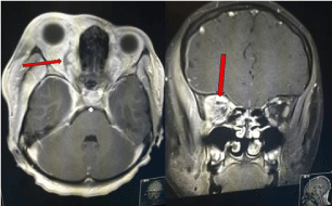
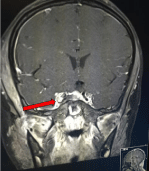
MRI of the head, neck, and orbit demonstrated a right retropharyngeal abscess 2.5 cm x 6 mm with a necrotic right retropharyngeal lymph node. There was right internal carotid artery vasospasm without thrombus or dissection, a dilated right superior ophthalmic vein (Figure 2) with a 5 mm thrombus in the right jugular bulb, and cavernous sinus thrombosis (Figure 3). IV Ceftriaxone and Meropenem were initiated. Surgical intervention was deferred given the extent of the thrombosis. Anticoagulation was also started.

Figure 2: MRI imaging, showing dilation of the right superior ophthalmic vein.

Figure 3: Right cavernous sinus thrombosis.
Her vision remained 20/20 throughout her hospital course. On hospital day 2, a perforated right otitis media was noted. Her ear drainage grew Methicillin Sensitive Staphylococcus Aureus (MSSA) for which otic ciprofloxacin and dexamethasone were added to her systemic antibiotics. On hospital day 3, IOP elevated to 32 on the right eye, at which point Cosopt was started. Blood cultures were eventually negative. A thrombophilia work-up was negative.
Eventually, her chemosis had resolved and extraocular movements returned to normal. Repeat MRI 6 days after initial hospitalization, showed improvement in swelling and thrombus. IV antibiotics were switched to meropenem and nafcillin on discharge based on resulting antibiotic sensitivities.
At 1 month follow up, she remained with 20/20 vision in both eyes, improved exophthalmos with Hertel measurements of 15 mm right eye, 13 mm left eye, base 96 mm. She had trace swelling of the right eyelid, and full extraocular movements bilaterally. MRI done 3 months post discharge revealed resolution of swelling and thrombus.
Discussion
Lemierre’s syndrome consists of a primary oropharyngeal infection, that leads to internal jugular vein thrombophlebitis and septic emboli, which cause hematologic spread and septicemia [1]. Fusobacterium necrophorum is the classic causative agent, though other bacteria include Peptostreptococcus, Group B and C. Streptococcus, Staphylococcus, and Enterococcus species [2]. The primary infection is usually pharyngitis in an estimated 87% of patients, but can also result from mastoiditis, tooth infections, as well as parotitis, skin infections and sinusitis [3]. In Lemierre’s original publication in Lancet, mortality rate of 18 out of 20 cases [1]. With the advent of intravenous antibiotics, the mortality rate has improved to at least 17% [4]. Classic symptoms include fever, oropharyngeal pain, neck swelling, arthralgia and pulmonary symptoms [4]. Typically, LD affects young adults, with a median age of 19, and 89% between 10 to 35 years of age [5].
Intracranial complications of Lemierre’s have been reported. The risk of developing intracranial involvement is significantly higher with otogenic (65.5%) vs postanginal (6.2%) Fusobacterium [5]. There are a small number of case reports of prior presentation of Lemierre’s syndrome associated with orbital involvement [6- 12]. Though Lemierre’s syndrome manifests with eye symptoms in only 5% of cases [2], loss of vision has been reported in case reports [11,12]. In this particular case report, the primary origin of infection was likely otogenic.
Anticoagulation remains a source of controversy for the management of LD. Internal jugular vein thrombosis is one typical component of LD. Arguments for anticoagulation include quicker improvement of bacteremia and thrombophlebitis [13]. Proposed indications include cerebral infarcts or sinus venous thrombosis [14] while others suggest anticoagulation only for retrograde progression of disease to the cavernous sinus [15] However, given the propensity for LD to be exacerbated by septic emboli, anticoagulation may aggravate the infection [4].
Lemierre’s syndrome, though primarily an oropharyngeal disease, can have systemic involvement. Though historically the disease had a high mortality rate, prognosis is significantly improved with current diagnostic abilities, systemic antibiotic therapy and close monitoring.
References
- Lemierre A. On certain septicaemias due to anaerobic organisms. Lancet. 1936; 227: 701-703.
- Karkos PD, Asrani S, Karkos CD, Leong SC, Theochari EG, Alexopoulou TD, et al. Lemierre’s syndrome: a systematic review. Laryngoscope. 2009; 119; 1552-1559.
- Chirinos JA, Lichtstein DM, Garcia J, Tamariz LJ. The evolution of Lemierre syndrome: report of 2 cases and review of the literature. Medicine (Baltimore). 2002; 81: 458-465.
- Hagelskjaerkristensen L, Prag J. Human necrobacillosis, with emphasis on Lemierre’s syndrome. Clin Infect Dis. 2000; 31: 524-532.
- Riordan T. Human infection with Fusobacterium necrophorum (Necrobacillosis), with a focus on Lemierre’s syndrome. ClinMicrobiol Rev. 2007; 20: 622-659.
- Figueras Nadal C, Creus A, Beatobe S, Moraga F, Pujol M, Vazquez E. Lemierre syndrome in a previously healthy young girl. ActaPaediatr. 2003; 92: 631-633.
- Hama Y, Koga M, Fujinami J, Asayama S, Toyoda K. Slowly progressive Lemierre’s syndrome with orbital pain and exophthalmos. J Infect Chemother. 2016; 22: 58-60
- Kahn JB, Baharestani S, Beck HC, et al. Orbital dissemination of Lemierre syndrome from gram-positive septic emboli. OphthalPlastReconstr Surg. 2011; 27: 67-68.
- Ballester DG, Moreno-sánchez M, González-garcía R, Gil FM. Lemierre syndrome: headache and proptosis as unusual presentation of dental infection by Gemellamorbillorum. Br J Oral Maxillofac Surg. 2016; 54: 842- 844.
- Shibuya K, Igarashi S, Sato T, Shinbo J, Sato A, Yamazaki M. [Case of Lemierre syndrome associated with infectious cavernous sinus thrombosis and septic meningitis]. RinshoShinkeigaku. 2012; 52: 782-785.
- Stauffer C, Josiah AF, Fortes M, Menaker J, Cole JW. Lemierre syndrome secondary to community-acquired methicillin-resistant Staphylococcus aureus infection associated with cavernous sinus thromboses. J Emerg Med. 2013; 44: 177-182.
- Akiyama K, Karaki M, Samukawa Y, Mori N. Blindness caused by septic superior ophthalmic vein thrombosis in a Lemierre Syndrome variant. AurisNasus Larynx. 2013; 40: 493-436.
- Ramirez S, Hild TG, Rudolph CN, et al. Increased diagnosis of Lemierre syndrome and other Fusobacterium necrophorum infections at a Children’s Hospital. Pediatrics. 2003; 112: 380.
- Bentham JR, Pollard AJ, Milford CA, Anslow P, Pike MG. Cerebral infarct and meningitis secondary to Lemierre’s syndrome. Pediatr Neurol. 2004; 30: 281-283.
- Riordan T, Wilson M. Lemierre’s syndrome: more than a historical curiosa. Postgrad Med J. 2004; 80: 328-333.