
Special Article - Conjunctivitis : Clinical Cases and Images
Austin J Clin Ophthalmol. 2016; 3(3): 1074.
Conjunctival Lipoma
Rene Hernan Parada Vasquez*
Department of Anterior Segment, Institute of Vision, Hospital la Carlota, Mexico
*Corresponding author: Rene Hernan Parada Vasquez, Department of Anterior Segment, Institute of Vision, Hospital la Carlota, Montemorelos, Nuevo Leon, Mexico
Received: December 05, 2016; Accepted: December 12, 2016; Published: December 14, 2016
Clinical Image
A 24-year-old female patient presented a slight bulging of the bulbar conjunctiva in right eye, about 5 mm. Outside the corneoscleral limbus, nearly to the outer cantus. The appearance of the massis cystic, mobile, giving clinical impression of conjunctival dermoid cyst (Figure 1A). Orbit CT scan with contrast was sent showing: Lipoma.
It proceeds to perform surgical removal of the cyst. It is sent to pathology, showing: Macroscopic: Tissuebiopsy 1.5x1x0.8 cm. (Figure 1B). Microscopic: Lesion formed by a dipocytes that are distributed as lobular and have fine fibrovascular trabeculae through them, as well as some areas of fibrosis. Diagnosis: Fibrolipoma.
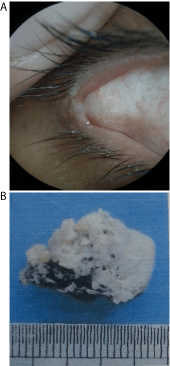
    Figure 1:  Conjunctival dermoid cyst.
A: Appearance of the lesion in right eye.
B: Conjunctival tissue biopsy, showing Fibrolipoma.