Research Article
Austin J Clin Ophthalmol. 2017; 4(1): 1078.
Measurement of Retinal Foveal Thickness and Foveal Volume during Pregnancy in Saudi Females Using Optical Coherence Tomography
Fahmy R1,2, Almatrafi M1 and Aldarwesh A1*
¹King Saud University, Department of Optometry and Vision Sciences, Riyadh, Saudi Arabia
²Department of Ophthalmology, Faculty of Medicine, Cairo University, Egypt
*Corresponding author: Aldarwesh AQ, Department of Optometry and Visual Sciences, King Saud University, P.O. Box 89885, Riyadh 11692, Saudi Arabia
Received: May 19, 2017; Accepted: June 12, 2017; Published: June 19, 2017
Abstract
Purpose: To evaluate whether pregnancy have an effect on the foveal thickness (FT) and volume (FV) in Saudi women.
Methods: This study involved 120 pregnant women (age range: 19-37 years) in the first, second and third trimester as the study groups and compared to 40 healthy non-pregnant women with similar age range.The assessment of FT and FV in each group was done using the optical coherence tomography (OCT).Statistics such as the mean, percentiles and range were used. A P-value of 0.05 or less was set to test significance.
Results: Average foveal thickness and foveal volume were significantly larger (P=0.05) in the first, second and third trimesters compared to controls. The mean FT was 226.57±23.3 μm, 231.37±19.21 μm, 240.3±21.76 μmin group 2,3, and 4 respectively compared to control mean of 214.2±19.73μm. Mean FV was 0.259±0.07 mm3, 0.241±0.05 mm3, 0.300±0.11 mm3 in group 2,3, and 4 respectively compared to control mean of 0.169±0.015 mm3. The 3rd trimester was associated with significantly (P=0.05) higher FV compared to the 2nd trimester.
Conclusion: The changes associated with pregnancy such as the increase of body fluids, can cause significant alteration in FT and FV. Although such alteration may not affect the quality of vision in healthy subjects, pregnant women with conditions that may cause fluid retention, or those with pre-existing systemic or eye disease or who are predisposed to risk factors should be monitored during pregnancy to avoid unexpected complications.
Keywords: Foveal thickness; Foveal volume; Pregnancy; Optical coherence tomography
Introduction
Pregnancy is associated with many physiological and physical changes that are attributed to the shift in hormonal levels, mainly the estrogen and progesterone during the reproductive phases of the first, second and third trimesters. These include gastrointestinal complaints, breast tenderness, fatigue, dizziness, gaining weight, frequent urination and pregnancy mask [1]. Moreover, the estrogen and progesterone were related to somatopsychologic disturbances in pregnancy as early as the first trimester [2,3].
The pregnancy imposes an effect on each organ system of the body, and the eye is no exception. Pregnancy-induced ocular changes include reduction in the intraocular pressure (IOP) [4], unilateral ptosis [5], dry eye [4] and alteration in lens and corneal curvature6,7 have been observed and attributed to fluid and hormonal changes [6- 8].
The ophthalmic changes could start as early as the first trimester and recovery to the pre-pregnancy levels could take weeks to months post-partum [6].
In pregnancy, plasma volume expands to support the uteroplacental blood flow to the fetus. The increase in total body water can be detected early in the pregnancy and continue till the delivery [9]. Pregnancy-induced corneal thickness due to edema has been reported [7]. An alteration in the refractive error and transient loss of accommodation may follow [7]. Fluid retention during pregnancy may affect other ocular tissue such as the choroid, increasing its thickness in the second or third trimesters [10,11]. Subfovealchoroidal areas were also found to be of higher thickness in healthy pregnant subjects [12,13]. Theses changes were detected using optical coherence tomography (OCT) [10-12].
On the other hand, pregnancy may exacerbate retinal diseases such as diabetic retinopathy (DR). Pregnant women with diabetes have a risk of developing either non-proliferative or proliferative DR [14]. One suggestion to the increased risk of DR during pregnancy is the increase in retinal blood flow and hyper-perfusion inducedstress on the compromised retina [15]. It has also been found that progesterone may influence the release of vascular endothelial growth factor (VEGF), a well-known angiogenic factor that exacerbates DR [16]. As the macular edema may develop or worsen during pregnancy, early detection is essential for the management and prevention of further complications.
Clinical examination involves the use of OCT, a non-invasive procedure, which gives high resolution cross-sectional, imaging that, enables the clinician to scan accurately, the subclinical changes in the retina. The measurement of central foveal thickness correlates with the visual acuity and provides a mean of monitoring the macula in different conditions [17]. In this study, we used OCT to quantify foveal thickness and volume in healthy pregnant women during the first, second and third trimesters and to assess whether pregnancyinduced foveal changes contribute to any visual disturbances in healthy subjects.
Methods
Study population and examination
All pregnant women were recruited from the gynecology and obstetrics department and ophthalmology unit in King Khalid University between March and May 2014.
This study included 120 healthy pregnant women (120 eyes) and 40 healthy non-pregnant women (40 eyes). Group 1 (control group) included 40 healthy non-pregnant women (40 eyes), Group 2 consisted of 40 healthy pregnant women (40 eyes) in first trimester; group 3 included 40 healthy pregnant women (40 eyes) in the second trimester; and group 4 included 40 healthy pregnant women (40 eyes) in third trimester.
Exclusion criteria for all participants included any history of diabetes mellitus, glaucoma, any corneal and lens pathologies preventing ocular fundus examination, laser therapy, and refractive error greater than 0.50 or less than -0.50 dioptres.
All participants underwent full ophthalmic examination in the form of: visual acuity test using E chart, auto refraction KR.8800, and complete slit-lamp examination of the anterior and posterior segment of the eye.
The values of FT and FV were measured by OCT RTVue model –RT100 (optovueInc, Fremont, CA) after mydriasis with tropicamide drop 1%. Three measurements were taken for the right eye and used for statistical analysis. The 6 macular map was used to evaluate the values of FT and FV. All measurements were carried out by same operator on all subjects.
Statistical analysis
The results were analyzed using Graphpad prism software version 7 (GraphPad Software, Inc., La Jolla, USA) and variables are presented as mean ± SD. D’Agostino & Pearson test was used to assess the normality of the data. One- way analysis of variance (ANOVA) was used to compare datasets. The level of significance was set at P value of 0.05 or less.
Ethical consideration
The protocol of the study was explained to each participant at the time of recruitment and informed consent was obtained according to the Declaration of Helsinki. The research was approved by the research ethical committee at King Khalid University Hospital (KKUH).
Results
The study included 120 eyes of 120 healthy pregnant women. The control group consisted of 40 eyes of 40 healthy non-pregnant women. There was no variation in age between the four groups. The mean age was: 30.4±4.1, 29.1±4.7, 28.03±5.4 and 27.4±4.6 years, in group 1,2,3 and 4 respectively. Table 1 shows the results of mean age, FT, and FV values in all participants.
    Group 1
    Group 2
    Group 3
    Group 4


    Age (years)
      Mean± SD
    30.4±4.1
    29.1±4.7
    28.03±5.4
    27.4±4.6


    FT (μm)


    Mean± SD
    214.2±19.7
    226.6±23.3
    231.4±19.2
    240.3±21.8


    5th Percentile
    175.2
    188.7
    200.5
    208.1


    Median
    216.5
    224.5
    225
    237.5


    95th Percentile
    224.9
    268.9
    274.5
    290


    FV (mm3)


    Mean± SD
    0.169±0.0158
    0.259±0.0706
    0.241±0.0515
    0.300±0.111


    5th Percentile
    0.14
    0.2
    0.16
    0.14


    Median
    0.17
    0.28
    0.23
    0.31


    95th Percentile
    0.19
    0.26
    0.24
    0.30


    FT:    Foveal Thickness; FV: Foveal Volume; SD: Standard Deviation.
Table 1: Mean age, FT and FV values of the groups.
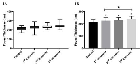
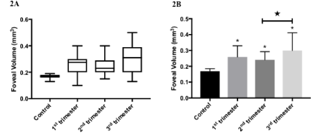
Data from this study showed that FT and FV values were increased significantly (P=0.05) during the pregnancy as early as the first trimester (Figure 1B and 2B). The mean FT was 226.57±23.3 μm, 231.37±19.21 μm, 240.3±21.76 μmin group 2,3, and 4 respectively compared to control mean of 214.2±19.73 μm. Mean FV was 0.259±0.07 mm3, 0.241±0.05 mm3, 0.300±0.11 mm3 in group 2,3, and 4 respectively compared to control mean of 0.169±0.015 mm3. Moreover, the mean FT was altered significantly (P=0.05) in the third trimester as compared to the control and the first trimester (Figure 1B). Similarly, the mean of the FV was significantly higher (P=0.05) compared to the second trimester (Figure 2B). No correlation was found between the FT and FV, the age and trimesters in all groups.

    Figure 1:  Foveal thickness (FT) measurement (μm) (Right eye, n=40).
1A: The box and Whisker’s plot showing the distribution of average, minimum
and maximum values of FT by the pregnancy trimester compared to control.
1B: The average FT of pregnant women in the first, second and third trimester
to compare to healthy non-pregnant control women.
*Indicates significant difference compared to control,
?Indicates significant difference between the third trimester compared to
the first trimester.

    Figure 2:  Foveal volume (FV) measurement (mm3) (Right eye, n=40)
1A: The box and Whisker’s plot showing the distribution of average, minimum
and maximum values of FV by the pregnancy trimester compared to control.
1B: The average FV of pregnant women in the first, second and third trimester
to compare to healthy non-pregnant control women.
*Indicates significant difference compared to control,
?Indicates significant difference between the third trimester compared to the
second trimester.
Discussion
The assessment of foveal thickness and volume are important for the diagnosis, treatment, and follow-up of retinal diseases such as diabetic retinopathy, age-related macular degeneration and glaucoma. In this study, OCT was utilized to detect changes to the fovea during pregnancy trimesters compared to healthy non-pregnant women. Several studies have shown alteration in the choroidal thickness during pregnancy especially in the third trimester [10-13,18,19]. The affected regions include the central subfield, inferior inner macular choroid, temporal, nasal and sub-foveal areas [12,19]. On the other hand, others reported that pregnancy has no effect on the choroidal thickness and it is comparable to healthy non-pregnant women [20,21].
The current results showed that pregnancy could alter significantly the FT and FV as compared to healthy non-pregnant values. This is in accordance with Demir et al., (2011) [22] findings in which women in their third trimester had higher mean values for the foveal and parafoveal thickness. One may expect that the third trimester is associated with more changes compared to earlier gestational periods; our study shows that earlier changes can also be detected in the first and second trimester using OCT. These were significantly higher in our sample. Similarly, there was a significant 53% and 77% increase in the FV in the first and third trimester respectively as compared to control. This is in parallel with Cankaya et al., (2013) [23] who found that total macular volume and FT were increased in the first, second and third trimesters in healthy women. As no pathological findings were found, this could be explained by the expansion of plasma volume in pregnancy, which is reported to increase by 45% [9,24]. One of the main limitations of the current study was the small sample size. In addition, it is worth monitoring one group from the first trimester till post-partum and monitor the time needed for the FT and FV values to return to the baseline.
In conclusion, pregnancy can result in an alteration to different ocular structures without visual complaints. However, routine examination of the retina using OCT is recommended for detecting early signs of macular edema especially if the patients are predisposed to any risk factors for developing hypertension, preeclampsia, diabetes or any other conditions that affect the retina.
Acknowledgements
This research project was supported by a grant from the “Research Center of the Female Scientific and Medical Colleges”, Deanship of Scientific Research, King Saud University.
References
- Jadotte YT, Schwartz RA. Melasma: insights and perspectives. Acta Dermatovenerol Croat. 2010; 18: 124-129.
- Polson JW, Paton JFR, Wolf AR. Altered autonomic function in programmed hypertension following gestational administration of glucocorticoid. Auton Neurosci. 2007; 135: 147.
- Weinstock M. The long-term behavioural consequences of prenatal stress. Neurosci Biobehav Rev. 2008; 32: 1073-1086.
- Ibraheem WA, Ibraheem AB, Tjani AM, Oladejo S, Adepoju S, Folohunso B. Tear Film Functions and Intraocular Pressure Changes in Pregnancy. Afr J Reprod Health. 2015; 19: 118-122.
- Sanke RF. Blepharoptosis as a complication of pregnancy. Ann Ophthalmol. 1984; 16: 720-722.
- Chawla S, Chaudhary T, Aggarwal S, Maiti GD, Jaiswal K, Yadav J. Ophthalmic considerations in pregnancy. Med J Armed Forces India. 2013; 69: 278-284.
- Park SB, Lindahl KJ, Temnycky GO, Aquavella JV. The effect of pregnancy on corneal curvature. CLAOJ. 1992; 18: 256-259.
- Yenerel, NM, Kucumen RB. Pregnancy and the Eye. Turk J Ophthalmol. 2015; 45: 213-219.
- Bernstein IM, Ziegler W, Badger GJ. Plasma volume expansion in early pregnancy. Obstet Gynecol. 2001; 97: 669-672.
- Dadaci Z, Alptekin H, OncelAcir N, Borazan M. Changes in choroidal thickness during pregnancy detected by enhanced depth imaging optical coherence tomography. Br J Ophthalmol. 2015; 99: 1255-1259.
- Goktas S, Basaran A, Sakarya Y, Ozcimen M, Kucukaydin Z, Sakarya, R, et al. Measurement of choroid thickness in pregnant women using enhanced depth imaging optical coherence tomography. Arq Bras Oftalmol. 2014; 77: 148-151.
- Sayin N, Kara N, Pirhan D, Vural A, ArazErsan HB, Tekirdag AI, et al. Subfovealchoroidal thickness in preeclampsia: comparison with normal pregnant and nonpregnant women. Semin Ophthalmol. 2014; 29: 11-17.
- Kara N, Sayin N, Pirhan D, Vural AD, Araz-Ersan HB, Tekirdag AI, et al. Evaluation of subfovealchoroidal thickness in pregnant women using enhanced depth imaging optical coherence tomography. Curr Eye Res. 2014; 39: 642-647.
- Sheth BP. Does pregnancy accelerate the rate of progression of diabetic retinopathy?: an update. Curr Diab Rep. 2008; 8: 270-273.
- Four risk factors for severe visual loss in diabetic retinopathy. The third report from the Diabetic Retinopathy Study. The Diabetic Retinopathy Study Research Group. Arch Ophthalmol. 1979; 97: 654-655.
- Swiatek-De Lange M, Stampfl A, Hauck SM, Zischka H, Gloeckner CJ, Deeg CA, et al. Membrane-initiated effects of progesterone on calcium dependent signaling and activation of VEGF gene expression in retinal glial cells. Glia. 2007; 55: 1061-1073.
- Hee MR, Puliafito CA, Wong C, Duker JS, Reichel E, Rutledge B, et al. Quantitative assessment of macular edema with optical coherence tomography. Arch Ophthalmol. 1995; 113: 1019-1029.
- Atas M, Acmaz G, Aksoy H, Demircan S, Atas F, Gulhan A, et al. Evaluation of the macula, retinal nerve fiber layer and choroid in preeclampsia, healthy pregnant and healthy non-pregnant women using spectral-domain optical coherence tomography. Hypertens Pregnancy. 2014; 33: 299-310.
- Rothwell RT, Meira DM, Oliveira MA, Ribeiro LF, Fonseca SL. Evaluation of Choroidal Thickness and Volume during the Third Trimester of Pregnancy using Enhanced Depth Imaging Optical Coherence Tomography: A Pilot Study. J Clin Diagn Res. 2015; 9: NC08-11.
- Kim JW, Park MH, Kim YJ, Kim YT. Comparison of subfovealchoroidal thickness in healthy pregnancy and pre-eclampsia. Eye (Lond). 2016; 30: 349-354.
- Takahashi J, Kado M, Mizumoto K, Igarashi S, Kojo T. Choroidal thickness in pregnant women measured by enhanced depth imaging optical coherence tomography. Jpn J Ophthalmol. 2013; 57: 435-439.
- Demir M, Oba E, Can E, Odabasi M, Tiryaki S, Ozdal E, et al. Foveal and parafoveal retinal thickness in healthy pregnant women in their last trimester. ClinOphthalmol. 2011; 5: 1397–1400.
- Cankaya C, Bozkurt M, Ulutas O. Total macular volume and foveal retinal thickness alterations in healthy pregnant women. SeminOphthalmol. 2013; 28: 103-111.
- Faupel-Badger JM, Hsieh CC, Troisi R, Lagiou P, Potischman N. Plasma volume expansion in pregnancy: implications for biomarkers in population studies. Cancer Epidemiol Biomarkers Prev. 2007; 16: 1720-1723.